. 2007;35(6):2003-12.
doi: 10.1093/nar/gkm063. Epub 2007 Mar 6.

Tat competes with HEXIM1 to increase the active pool of P-TEFb for HIV-1 transcription

Affiliations

Tat competes with HEXIM1 to increase the active pool of P-TEFb for HIV-1 transcription

Matjaz Barboric et al. Nucleic Acids Res. 2007.

Abstract

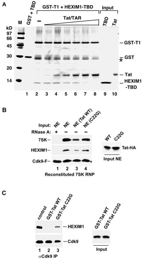
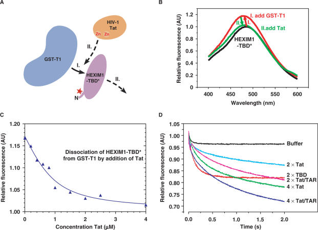
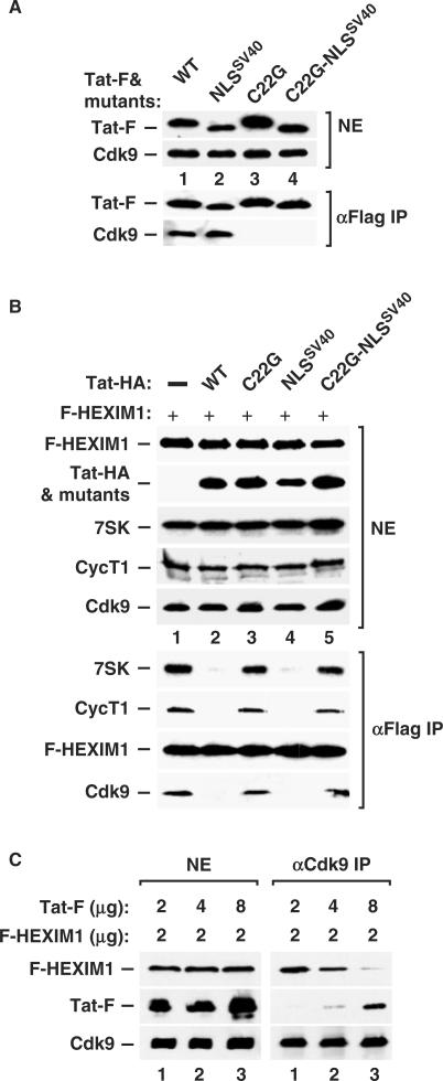
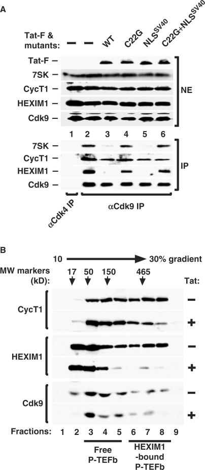
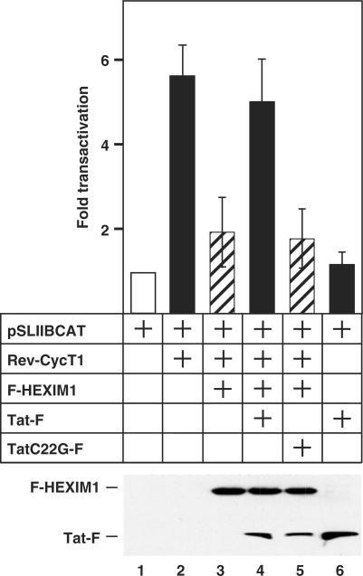
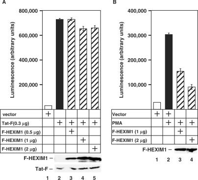
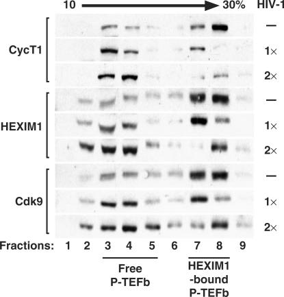
Human immunodeficiency virus type 1 (HIV-1) transcriptional transactivator (Tat) recruits the positive transcription elongation factor b (P-TEFb) to the viral promoter. Consisting of cyclin dependent kinase 9 (Cdk9) and cyclin T1, P-TEFb phosphorylates RNA polymerase II and the negative transcription elongation factor to stimulate the elongation of HIV-1 genes. A major fraction of nuclear P-TEFb is sequestered into a transcriptionally inactive 7SK small nuclear ribonucleoprotein (snRNP) by the coordinated actions of the 7SK small nuclear RNA (snRNA) and hexamethylene bisacetamide (HMBA) induced protein 1 (HEXIM1). In this study, we demonstrate that Tat prevents the formation of and also releases P-TEFb from the 7SK snRNP in vitro and in vivo. This ability of Tat depends on the integrity of its N-terminal activation domain and stems from the high affinity interaction between Tat and cyclin T1, which allows Tat to directly displace HEXIM1 from cyclin T1. Furthermore, we find that in contrast to the Tat-independent activation of the HIV-1 promoter, Tat-dependent HIV-1 transcription is largely insensitive to the inhibition by HEXIM1. Finally, primary blood lymphocytes display a reduced amount of the endogenous 7SK snRNP upon HIV-1 infection. All these data are consistent with the model that Tat not only recruits but also increases the active pool of P-TEFb for efficient HIV-1 transcription.


Figures

Figure 1.
Figure 1.
Tat disrupts the HEXIM1-P-TEFb interaction in vitro. (A) Competition between HEXIM1-TBD and Tat for binding to GST-T1. GST alone (lane 1) or GST-T1 (lanes 2–8) was incubated with HEXIM1-TBD. Increasing amounts of Tat and TAR (kept at an 1:1 molar ratio) were added to the reactions in lanes 3–8. The bound proteins were detected in a Coomassie–stained SDS-gel. Lanes 9 and 10 represent 40% of the input proteins. The bands denoted with a star (*) represent degradation products of GST-T1. (B) Tat suppresses the in vitro reconstitution of 7SK snRNP. To the immobilized P-TEFb (through Cdk9-F), RNase A-treated or untreated HeLa NE (lanes 1 and 2) or NE containing the wild-type or mutant Tat-HA proteins (lanes 3 and 4) was added as indicated. The amounts of 7SK and HEXIM1 bound to P-TEFb (left panel) and the levels of Tat-HA proteins in NE (right panel) are determined by northern and western blotting. (C) Tat disrupts the 7SK snRNP in vitro. 7SK snRNP immobilized on anti-Cdk9 beads were incubated with the wild-type or mutant C22G GST-Tat chimeras as indicated. The levels of Cdk9-bound HEXIM1 (left panel) and the input GST-Tat chimeras (right panel) are determined by western blotting.
Figure 2.
Figure 2.
Tat displaces HEXIM1-TBD from CycT1 due to its higher affinity for CycT1. (A) A schematic diagram of the main steps of the fluorescence competition assay. GST-T1 was first added to HEXIM1-TBD* (step I), followed by the addition of Tat (step II). (B) Fluorescence emission spectra of the two-step titrations as diagrammed in A. To 1.0 µM HEXIM1-TBD* (black curve), 1.0 µM of GST-T1 was added (red), followed by an excess of 5.0 µM Tat (green). (C) Dissociation of the HEXIM1-TBD* from GST-T1 by increasing concentrations of Tat. The displacement of HEXIM1-TBD* from GST-T1 by Tat was observed by equilibrium fluorescence titration. To a preformed complex of 2.0 µM HEXIM-TBD* and 1.0 µM GST-T1, Tat was added at concentrations from 0.2–4 µM. (D) Kinetics of the Tat- or Tat/TAR-mediated displacement of HEXIM1-TBD* from GST-T1 as measured by stopped flow techniques. To a pre-equilibrated solution of 5 µM GST-T1 and 5 µM dimeric HEXIM1-TBD*, Tat or Tat/TAR was injected at a concentration of 10 (2×) or 20 µM (4×) to displace HEXIM1-TBD* over the indicated time periods (in seconds). The displacement caused by buffer alone or 10 µM (2×) unlabeled HEXIM1-TBD (labeled as TBD) is also presented as a comparison.
Figure 3.
Figure 3.
Tat prevents HEXIM1 from binding to P-TEFb in vivo. (A) Western analyses of the levels of Cdk9 as well as the indicated wild-type or mutant Tat-F proteins in NEs of transfected HeLa cells (upper panels; all plasmids transfected at 2 µg per 15-cm dish) and the levels of Cdk9 associated with Tat-F in the anti-FLAG immunoprecipitates (lower panels). (B) HeLa cells were co-transfected with appropriate empty vectors (−) or vectors coding for F-HEXIM1 (2 µg/15-cm dish) and various Tat-HA proteins (2 µg) as indicated (total 4 µg plasmid DNA per dish). The levels of F-HEXIM1, Tat-HA, 7SK, CycT1 and Cdk9 in NEs (top five panels) and the amounts of 7SK, CycT1 and Cdk9 bound to the immunoprecipitated F-HEXIM1 (bottom four panels) are examined by western and northern blotting. (C) HeLa cells were co-transfected with a constant amount (2 µg/15-cm dish) of the plasmid coding for F-HEXIM1 and increasing amounts (2, 4 and 8 µg) of the plasmid coding for wild-type Tat-F. The levels of F-HEXIM1, Tat-F and Cdk9 proteins in NEs of transfected cells as well as the amounts of F-HEXIM1 and Tat-F proteins bound to the immunoprecipitated Cdk9 are detected by western blotting and shown in the left and right panels, respectively.
Figure 4.
Figure 4.
Tat disrupts the endogenous 7SK snRNP to release P-TEFb. (A) NEs prepared from HeLa cells either untransfected (−) or transfected with the indicated plasmids (20 µg per 15–cm dish) encoding wild-type or mutant Tat-F proteins were subjected to anti-Cdk9 or, as a negative control, anti-Cdk4 immunoprecipitation. The levels of endogenous 7SK, CycT1 and HEXIM1 bound to Cdk9 as revealed by northern or western blotting are indicated (bottom panels). The upper panels show the levels of the endogenous 7SK, CycT1, HEXIM1 and Cdk9 as well as the various transfected Tat-F proteins in HeLa NEs. (B) NEs prepared from HeLa cells transfected with an empty vector (−) or the Tat-F-encoding plasmid (20 µg/15-cm dish) were subjected to glycerol gradient sedimentation analysis. The panels show the western detection of CycT1, HEXIM1 and Cdk9 in gradient fractions (left). Molecular size standards were analyzed in a parallel gradient and their positions indicated by arrows.
Figure 5.
Figure 5.
Tat reverses the HEXIM1-mediated inhibition of P-TEFb transcriptional activity. HeLa cells were co-transfected with the reporter plasmid pSLIIBCAT (0.2 μg) and the plasmids coding for the various effectors (Rev-CycT1: 0.6 μg; F-HEXIM1: 0.6 μg; Tat-F: 0.8 μg; TatC22G-F: 0.8 μg) as indicated. The chloramphenicol acetyltransferase (CAT) enzyme activities in cell lysates were measured and the error bars represent the mean +/− SD. Lower panel shows the expression levels of the co-transfected F-HEXIM1 and Tat-F as revealed by western blotting.
Figure 6.
Figure 6.
HEXIM1 inhibits Tat-independent but not Tat-dependent transcriptional activation from the HIV-1 LTR. (A,B) HL3T1 cells containing an integrated HIV-1 LTR-driven luciferase reporter construct were transfected with an empty vector or the plasmids encoding Tat-F and/or F-HEXIM1. Where indicated, the cells were also stimulated by PMA. Luciferase activities in cell lysates were measured and the error bars represent the mean +/− SD. Lower panels show the expression levels of the co-transfected F-HEXIM1 and Tat-F as revealed by western blotting.
Figure 7.
Figure 7.
HIV-1-infected PBLs contain a reduced amount of 7SK snRNP. Total cell lysates prepared from uninfected (−) or HIV-1-infected PBLs at two different viral concentrations (1× or 2×) were subjected to glycerol gradient sedimentation analysis in a 10–30% gradient. The panels show western detection of HEXIM1, Cdk9 and CycT1 in gradient fractions with the positions of the HEXIM1-bound P-TEFb and the 7SK/HEXIM1-free P-TEFb marked at the bottom. For HIV infection, 1× equals to 3.22 ng/ml and 2× to 6.32 ng/ml of HIV-1 p24 detected in the supernatants at day 5 post infection.

References

    1. Barboric M, Peterlin BM. A new paradigm in eukaryotic biology: HIV Tat and the control of transcriptional elongation. PLoS Biol. 2005;3:e76. - PMC - PubMed
    1. Price DH. P-TEFb, a cyclin-dependent kinase controlling elongation by RNA polymerase II. Mol. Cell Biol. 2000;20:2629–2634. - PMC - PubMed
    1. Yik JH, Chen R, Nishimura R, Jennings JL, Link AJ, Zhou Q. Inhibition of P-TEFb (CDK9/Cyclin T) kinase and RNA polymerase II transcription by the coordinated actions of HEXIM1 and 7SK snRNA. Mol Cell. 2003;12:971–982. - PubMed
    1. Michels AA, Nguyen VT, Fraldi A, Labas V, Edwards M, Bonnet F, Lania L, Bensaude O. MAQ1 and 7SK RNA interact with CDK9/cyclin T complexes in a transcription–dependent manner. Mol. Cell Biol. 2003;23:4859–4869. - PMC - PubMed
    1. Michels AA, Fraldi A, Li Q, Adamson TE, Bonnet F, Nguyen VT, Sedore SC, Price JP, Price DH, et al. Binding of the 7SK snRNA turns the HEXIM1 protein into a P-TEFb (CDK9/cyclin T) inhibitor. EMBO J. 2004;23:2608–2619. - PMC - PubMed

Publication types

MeSH terms