. 2007;35(6):1992-2002.
doi: 10.1093/nar/gkm041. Epub 2007 Mar 6.

USER friendly DNA engineering and cloning method by uracil excision

Affiliations

USER friendly DNA engineering and cloning method by uracil excision

Jurate Bitinaite et al. Nucleic Acids Res. 2007.

Abstract

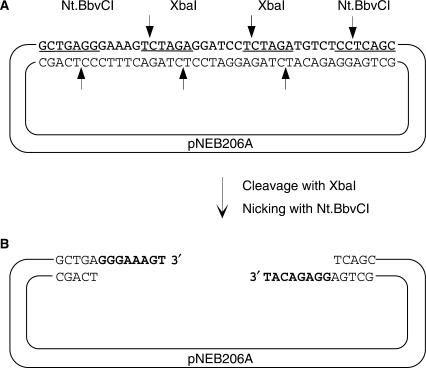
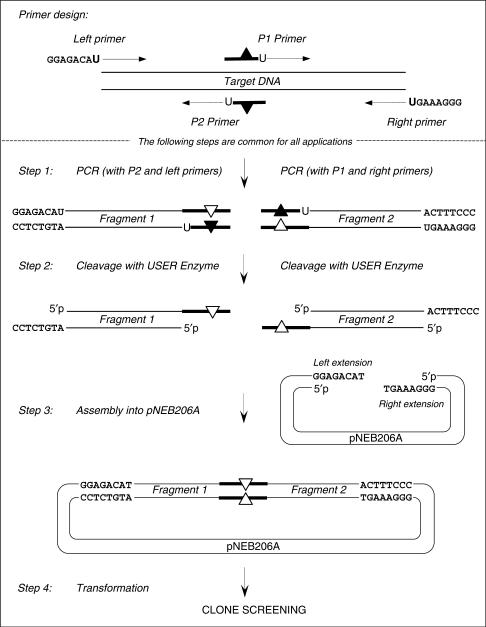
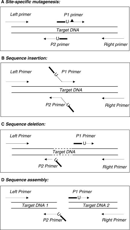
Here we report a PCR-based DNA engineering technique for seamless assembly of recombinant molecules from multiple components. We create cloning vector and target molecules flanked with compatible single-stranded (ss) extensions. The vector contains a cassette with two inversely oriented nicking endonuclease sites separated by restriction endonuclease site(s). The spacer sequences between the nicking and restriction sites are tailored to create ss extensions of custom sequence. The vector is then linearized by digestion with nicking and restriction endonucleases. To generate target molecules, a single deoxyuridine (dU) residue is placed 6-10 nt away from the 5'-end of each PCR primer. 5' of dU the primer sequence is compatible either with an ss extension on the vector or with the ss extension of the next-in-line PCR product. After amplification, the dU is excised from the PCR products with the USER enzyme leaving PCR products flanked by 3' ss extensions. When mixed together, the linearized vector and PCR products directionally assemble into a recombinant molecule through complementary ss extensions. By varying the design of the PCR primers, the protocol is easily adapted to perform one or more simultaneous DNA manipulations such as directional cloning, site-specific mutagenesis, sequence insertion or deletion and sequence assembly.


Figures

Figure 1.
Figure 1.
Schematic representation of the pNEB206A cloning vector. (A) pNEB206A vector was constructed by ligating a synthetic double-stranded cassette into the PacI and PmeI sites of pNEB193. Within the cassette, XbaI and Nt.BbvCI recognition sequences are underlined; cleavage sites are shown by arrows. (B) For USER friendly cloning, pNEB206A is double-digested with XbaI and Nt.BbvCI as described in the Materials and methods section to produce linearized vector flanked by 3′ single-stranded extensions on both ends.
Figure 2.
Figure 2.
Schematic description of the USER friendly DNA Engineering method. Lines with the arrowheads symbolize PCR primers. Boldface lines within the P1 and P2 primers and PCR products symbolize complementary overlapping sequences. Black triangles refer to the point mismatches in the primers and white triangles refer to the resulting mutations in the PCR products. dU is indicated by ‘U’. The specific sequences GGAGACAU in the left primer and GGGAAAGU in the right primer are designed for their compatibility with the vector pNEB206A sequence.
Figure 3.
Figure 3.
PCR primer design for various DNA manipulations. Only the primer design aspects of the indicated DNA manipulations are shown. The indicated DNA manipulations are completed by performing steps 1–4 shown in Figure 2. Lines with the arrowheads symbolize priming sequences in PCR primers. Boldface lines within primers symbolize overlapping sequences. dU is indicated by ‘U’ and flank the overlapping sequences on their 3′ side. (A) Site-specific mutagenesis. The desired sequence change, shown as a black triangle, is introduced downstream of dU into either of the overlapping primers P1 or P2. An alternative is shown in Figure 2 where sequence changes are introduced into the overlap sequence of both primers. (B) Nucleotide sequence insertion. Overlapping primers P1 and P2 precisely prime the template adjacent to the insertion site. The desired non-priming sequences are added to the 5′ ends of both primers (shown as angled lines). The overlapping sequence is created from the 5′ terminal 6–10 nucleotides of the insertion sequences (shown as the boldface angled lines), while the rest of the insertion sequence (shown as a dotted angled line) may be of any desired length. (C) Nucleotide sequence deletion. The overlapping primers P1 and P2 prime distant locations precisely adjacent to the targeted deletion region (shown as a dotted line). To create the overlapping sequence, the 5′ end of one primer (P2) is supplemented by a non-priming sequence complementary to the 5′ end sequence of the other primer (P1). (D) Sequence assembly. The overlapping primers, P1 and P2, prime different DNA templates. A complementary copy of the 5′ terminal sequence of one primer (P1) is added to the 5′ terminus of the other primer (P2) to create the overlapping sequence necessary for joining two independent DNAs.
Figure 4.
Figure 4.
USER enzyme activity assay. A 34-mer oligonucleotide duplex (10 pmol) containing a single dU paired with a deoxyadenine (Table 1) was incubated with a series of EndoVIII and UDG enzyme mixtures for 15 min at 37°C in a 10 μl of T4 DNA ligase reaction buffer (50 mM Tris-HCl pH 7.5, 10 mM MgCl2, 10 mM DTT, 1 mM ATP, 20 μg/ml BSA). The reactions were quenched by the addition of 10 μl of 95% formamide, 0.1% xylene cyanol, 0.1% bromophenol blue, 10 mM EDTA, pH 11, and the reaction products were analyzedon a 15% TBE-Urea denaturing gel. S, 34-nt oligonucleotide substrate. P1 and P2, 15 nt and 18 nt cleavage products, respectively. Two product bands of differing size result from the fact that the uracil is not in the centre of the substrate. Lane 1—no enzyme added. Lane 2—reaction contains 0.2 units of UDG. Lane 3—reaction contains 256 ng of EndoVIII. Lanes 4–10, reaction contains 0.2 units of UDG and the amount of EndoVIII shown above the respective lanes.
Figure 5.
Figure 5.
Outline of the chloramphenicol (Cat) gene mutagenesis and assembly experiment. (A) PCR primer design strategy. Lines with the arrowheads symbolize priming sequences in the PCR primers. Left and Right cloning primers contain 5′ extensions compatible with the single-stranded extensions on the linearized pNEB206A vector. The overlapping primers P1/P2 and P3/P4 are selected in the vicinity of the targeted mutations. Asterisks designate the point mismatches in the primers compared to the template sequence. Overlapping primers are complementary to each other within the boxed sequences. The 3′ dT of the boxed sequence in the primer sequences is replaced by dU. (B) Schematic representation of six PCR products amplified using the indicated primers. Cycling conditions are described in Materials and Methods. The label, Cat926 etc. shows the PCR fragment size in bp. The ends of the PCR products are flanked by the compatible overlapping sequences with a single dU residue at the junction. (C) Four assembly reactions were carried out using the indicated PCR fragments to assemble a full-length Cat gene carrying the indicated phenotype into pNEB206A.
Figure 6.
Figure 6.
Rheoactivator gene engineering. (A) Electrophoretic analysis of the PCR products amplified using PfuCx DNA Polymerase. 5 μl of each PCR reaction corresponding either to the CMV promoter amplification products (Lanes 1–3) or to the Rheoactivator gene amplification products (Lanes 4–7) were loaded on a 1% agarose gel. Lane 1—PCR with primers R47 + R48 (product size 153 bp). Lane 2—PCR with primers R49 + R50 (product size 184 bp). Lane 3—PCR with primers R51 + R52 (product size 218 bp). Lane 4—PCR with primers R53 + R54 (product size 144 bp). Lane 5—PCR with primers R55 + R56 (product size 142 bp). Lane 6—PCR with primers R57 + R58 (product size 190 bp). Lane 7—PCR with primers R59 + R60 (product size 470 bp). Lane 8—50 μg of the linearized pNEB206A vector. Lane 9—control ligation of the USER-treated PCR fragments shown in Lanes 1–7. White arrow indicates the 1500-bp ligation product that represents the full-length CMVpromoter and Rheoactivator gene fusion. Lane M corresponds to the 2-log DNA ladder. (B) Clone screening by PCR amplification directly from 10 white colonies with primers R51 and R56 that will only amplify clones carrying a CMV promoter and Rheoactivator gene fusion. (C), Plasmid DNA from clones 1, 2, 4, 5, 7 and 8 were analyzed by restriction digestion with BbvCI restriction endonuclease.

References

    1. Kunkel TA. Rapid and efficient site-specific mutagenesis without phenotypic selection. Proc. Natl. Acad. Sci. U.S.A. 1985;82:488–492. - PMC - PubMed
    1. Hofer B, Kuhlein B. A high efficiency method for site-directed mutagenesis with any plasmid. Gene. 1989;84:153–157. - PubMed
    1. Taylor JW, Ott J, Eckstein F. The rapid generation of oligonucleotide-directed mutations at high frequency using phosphorothioate-modified DNA. Nucleic Acids Res. 1985;13:8764–8785. - PMC - PubMed
    1. Hashimoto-Gotoh T, Mizuno T, Ogasahara Y, Nakagawa M. An oligodeoxyribonucleotide-directed dual amber method for site-directed mutagenesis. Gene. 1995;152:271–275. - PubMed
    1. Deng WP, Nickoloff JA. Site-directed mutagenesis of virtually any plasmid by eliminating a unique site. Anal. Biochem. 1992;200:81–88. - PubMed

Publication types

MeSH terms