Review
. 2007 Mar 7;2(3):e266.
doi: 10.1371/journal.pone.0000266.

Plasmids and rickettsial evolution: insight from Rickettsia felis

Affiliations
Review

Plasmids and rickettsial evolution: insight from Rickettsia felis

Joseph J Gillespie et al. PLoS One. .

Abstract

Background: The genome sequence of Rickettsia felis revealed a number of rickettsial genetic anomalies that likely contribute not only to a large genome size relative to other rickettsiae, but also to phenotypic oddities that have confounded the categorization of R. felis as either typhus group (TG) or spotted fever group (SFG) rickettsiae. Most intriguing was the first report from rickettsiae of a conjugative plasmid (pRF) that contains 68 putative open reading frames, several of which are predicted to encode proteins with high similarity to conjugative machinery in other plasmid-containing bacteria.

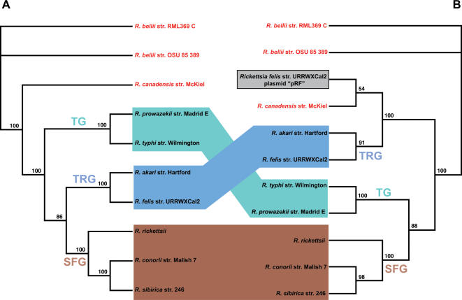
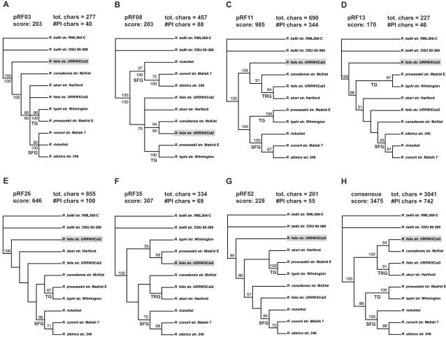
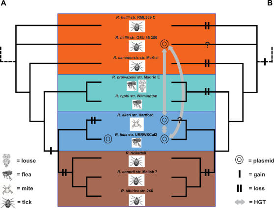
Methodology/principal findings: Using phylogeny estimation, we determined the mode of inheritance of pRF genes relative to conserved rickettsial chromosomal genes. Phylogenies of chromosomal genes were in agreement with other published rickettsial trees. However, phylogenies including pRF genes yielded different topologies and suggest a close relationship between pRF and ancestral group (AG) rickettsiae, including the recently completed genome of R. bellii str. RML369-C. This relatedness is further supported by the distribution of pRF genes across other rickettsiae, as 10 pRF genes (or inactive derivatives) also occur in AG (but not SFG) rickettsiae, with five of these genes characteristic of typical plasmids. Detailed characterization of pRF genes resulted in two novel findings: the identification of oriV and replication termination regions, and the likelihood that a second proposed plasmid, pRFdelta, is an artifact of the original genome assembly.

Conclusion/significance: Altogether, we propose a new rickettsial classification scheme with the addition of a fourth lineage, transitional group (TRG) rickettsiae, that is unique from TG and SFG rickettsiae and harbors genes from possible exchanges with AG rickettsiae via conjugation. We offer insight into the evolution of a plastic plasmid system in rickettsiae, including the role plasmids may have played in the acquirement of virulence traits in pathogenic strains, and the likely origin of plasmids within the rickettsial tree.


Conflict of interest statement

Competing Interests: The authors have declared that no competing interests exist.

Figures

Figure 1
Figure 1. Phylogeny estimation from analysis of fifteen R. felis proteins.
Phylogeny estimation under parsimony of fifteen R. felis proteins (hypothetical protein RF_0005, threonyl-tRNA synthetase, preprotein translocase SecA subunit, uncharacterized low-complexity protein RF_0864, pyruvate phosphate dikinase precursor, leucyl-tRNA synthetase, hypothetical protein RF_0556, NAD-specific glutamate dehydrogenase, DNA polymerase III alpha chain, O-antigen export system permease protein RfbA, thioredoxin, NADPH-dependent glutamate synthase beta chain and related oxidoreductases, putative TIM-barrel protein in nifR3 family, UDP-3-O-[3-hydroxymyristoyl] glucosamine, and zinc/manganese ABC transporter substrate binding protein TroA_c) from nine rickettsial species (Rickettsia bellii, R. canadensis, R. prowazekii, R. typhi, R. akari, R. felis, R. conorii, R. rickettsii, and R. sibirica) and two strains of Wolbachia. Branch support is from one million bootstrap replicates. Genome information was compiled from the PATRIC Website. * Total R. felis genome size: 1,485,148 bp = chromosome; 62,829 bp = pRF and 39,263 bp = pRFδ.
Figure 2
Figure 2. Comparison of phylogeny estimations from exclusively chromosomal proteins and proteins present on the chromosome and plasmids of R. felis.
(A) Estimated phylogeny of 21 exclusively chromosomal proteins from 10 rickettsial strains. (B) Estimated phylogeny of 10 proteins present on the chromosome and plasmids of R. felis. “Ancestral” (red) refers to primitive rickettsiae with no known potential for host virulence. TG (aquamarine) = typhus group, TRG (light blue) = transitional group and SFG (brown) = spotted fever group. TG and TRG boxes depict the major differences in tree topologies. The pRF genes are boxed and shaded. Results from both analyses of amino acids are from an exhaustive search under parsimony with branch support from one million bootstrap replications.
Figure 3
Figure 3. Individual phylogeny estimations for the seven pRF proteins used in the combined analysis of pRF.
(A, B) Majority rule consensus trees. (C–H) Strict consensus trees. All analyses were of amino acids from an exhaustive search under parsimony with branch support from one million bootstrap replications. Bootstrap values are placed above branches. Percentages of nodes recovered in majority rule consensus trees are shown below branches. Scores are tree lengths, with total characters and number of parsimony informative characters provided.
Figure 4
Figure 4. Characteristics and summary information of predicted origin of replication (oriV) of the pRF plasmid of Rickettsia felis.
(A) Schematic map of the pRF with shaded regions containing the putative oriV (right) and replication termination region (left). The region outlined in the dark dashed line depicts the portion of the plasmid missing in pRFδ (pRF15-pRF38). Grey boxes depict genes, with gene names described in Tables 1 and 2. Red lines depict coding strands, and yellow blocks depict areas of gene overlap. (B) AT-skew of pRF, with AT-skew (blue), cumulative AT-skew (red) and minimum AT-skew (orange). (C) CG-skew of pRF, with CG-skew (blue), cumulative CG-skew (red) and maximum CG-skew (orange). Plots generated and values computed with GenSkew (http://mips.gsf.de/services/analysis/genskew).
Figure 5
Figure 5. Comparison of two hypotheses for the evolution of plasmids in rickettsiae.
(A) The appearance of a plasmid system in R. felis (as a member of SFG rickettsiae) as recently suggested (Ogata et al., 2005b; Blanc et al. 2007). (B) Our hypothesis centered on the notion that the ancestor to all rickettsiae harbored a plasmid system with subsequent losses in the ancestors to the TG and SFG rickettsiae, and in R. canadensis and R. bellii str. RML369-C. Red = ancestral rickettsiae, Aquamarine = typhus group, light blue = transitional group, brown = true spotted fever group. Trees are from Figure 2A.

References

    1. Perlman SJ, Hunter MS, Zchori-Fein E. The emerging diversity of Rickettsia. Proc Biol Sci. 2006;273:2097–2106. - PMC - PubMed
    1. Hackstadt T. The biology of rickettsiae. Infect Agents & Disease. 1996;5:127–143. - PubMed
    1. Azad AF, Beard CB. Rickettsial pathogens and their arthropod vectors. Emerg Infect Dis. 1998;4:179–86. - PMC - PubMed
    1. Azad AF, Radulovic S. Pathogenic rickettsiae as bioterrorism agents. Ann N Y Acad Sci. 2003;990:734–738. - PubMed
    1. Weisburg WG, Dobson ME, Samuel JE, Dasch GA, Mallavia LP, et al. Phylogenetic diversity of the Rickettsiae. J Bacteriol. 1989;171:4202–4206. - PMC - PubMed

Publication types