Recovery of small-sized blood vessels in ischemic bone under static magnetic field
- PMID: 17342242
- PMCID: PMC1810364
- DOI: 10.1093/ecam/nel055
Recovery of small-sized blood vessels in ischemic bone under static magnetic field
Abstract
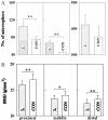
Effects of static magnetic field (SMF) on the vascularization in bone were evaluated using an ischemic bone model, where rat femoral artery was ligated. Magnetized and unmagnetized samarium-cobalt rods were implanted transcortically into the middle diaphysis of the ischemic femurs. Collateral circulation was evaluated by injection of microspheres into the abdominal aorta at the third week after ligation. It was found that the bone implanted with a magnetized rod showed a larger amount of trapped microspheres than that with an unmagnetized rod at the proximal and the distal region (P < 0.05 proximal region). There were no significant differences at the middle and the distal region. This tendency was similar to that of the bone mineral density in the SMF-exposed ischemic bone.
Figures


References
-
- Bassett CAL, Pawluk RJ, Becker RO. Effects of electrical current on bone in vivo. Nature. 1964;204:652–4. - PubMed
-
- Bassett CAL, Pawluk RJ, Pilla AA. Augmentation of bone repair by inductively coupled electromagnetic fields. Science. 1974;184:575–7. - PubMed
-
- Wiendl HJ, Strigl M. Clinical experiences in supplementary treatment of pseudarthroses using electromagnetic potentials. Fortschr Med. 1978;96:231–6. - PubMed
-
- Hanft JR, Goggin JP, Landsman A, Surprenant M. The role of combined magnetic field bone growth stimulation as an adjunct in the treatment of neuroarthropathy/Charcot joint: an expanded pilot study. J Foot Ankle Surg. 1998;37:510–5. discussion 550–1. - PubMed
-
- Bassett CAL, Mitchell SN, Gaston SR. Treatment of un-united tibial diaphyseal fractures with pulsing electromagnetic fields. J Bone Joint Surg. 1981;63A:511–23. - PubMed
LinkOut - more resources
Full Text Sources

