

Efficacy of static magnetic field for locomotor activity of experimental osteopenia
- PMID: 17342247
- PMCID: PMC1810356
- DOI: 10.1093/ecam/nel067
Efficacy of static magnetic field for locomotor activity of experimental osteopenia
Abstract
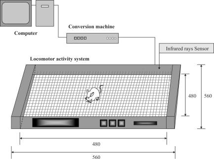
In order to examine the effectiveness of applying a static magnetic field (SMF) for increasing bone mineral density (BMD), we assessed the degree of osteopenia by dual-energy X-ray absorptiometry (DEXA), the metabolism measuring system, and histological examination of bone tissue in an ovariectomized (OVX) rat model. Thirty-six female Wistar rats (8 weeks old, 160-180 g) were divided into three groups. The rats in the OVX-M group were exposed to SMF for 12 weeks after ovariectomy. The ovariectomized rats in the OVX-D group were not exposed to SMF as a control. The rats in the normal group received neither ovariectomy nor exposure to SMF. Twelve-week exposure to SMF in the OVX-M group inhibited the reduction in BMD that was observed in the OVX-D group. Moreover, in the OVX rats, before exposure to SMF, there was no clear difference in the level of locomotor activity between the active and resting phases, and the pattern of locomotor activity was irregular. After exposure of OVX rats to SMF, the pattern of locomotor activity became diphasic with clear active and resting phases, as was observed in the normal group. In the OVX-M group, the continuity of the trabecular bone was maintained more favorably and bone mass was higher than the respective parameters in the OVX-D group. These results demonstrate that exposure to SMF increased the level of locomotor activity in OVX rats, thereby increasing BMD.
Figures


Similar articles
-
Effect of kami-kihi-to (jia-wei-gui-pi-tang) for experimental osteopenia.Am J Chin Med. 2005;33(1):41-8. doi: 10.1142/S0192415X05002643. Am J Chin Med. 2005. PMID: 15844832
-
The effects of nandrolone decanoate on bone mass and metabolism in ovariectomized rats with osteopenia.J Bone Miner Metab. 2000;18(5):258-63. doi: 10.1007/pl00010639. J Bone Miner Metab. 2000. PMID: 10959614
-
Recovery Effects of a 180 mT Static Magnetic Field on Bone Mineral Density of Osteoporotic Lumbar Vertebrae in Ovariectomized Rats.Evid Based Complement Alternat Med. 2011;2011:620984. doi: 10.1155/2011/620984. Epub 2010 Sep 28. Evid Based Complement Alternat Med. 2011. PMID: 20953437 Free PMC article.
-
Jumping exercise preserves bone mineral density and mechanical properties in osteopenic ovariectomized rats even following established osteopenia.Osteoporos Int. 2017 Apr;28(4):1461-1471. doi: 10.1007/s00198-017-3905-7. Epub 2017 Jan 26. Osteoporos Int. 2017. PMID: 28124728
-
Study on Application of Static Magnetic Field for Adjuvant Arthritis Rats.Evid Based Complement Alternat Med. 2004 Sep 1;1(2):187-191. doi: 10.1093/ecam/neh024. Evid Based Complement Alternat Med. 2004. PMID: 15480444 Free PMC article.
Cited by
-
Chronic Exposure to Static Magnetic Fields from Magnetic Resonance Imaging Devices Deserves Screening for Osteoporosis and Vitamin D Levels: A Rat Model.Int J Environ Res Public Health. 2015 Jul 30;12(8):8919-32. doi: 10.3390/ijerph120808919. Int J Environ Res Public Health. 2015. PMID: 26264009 Free PMC article.
-
Static magnetic fields in regenerative medicine.APL Bioeng. 2024 Mar 13;8(1):011503. doi: 10.1063/5.0191803. eCollection 2024 Mar. APL Bioeng. 2024. PMID: 38486824 Free PMC article. Review.
-
Physical stimulations and their osteogenesis-inducing mechanisms.Int J Bioprint. 2018 Jun 11;4(2):138. doi: 10.18063/IJB.v4i2.138. eCollection 2018. Int J Bioprint. 2018. PMID: 33102916 Free PMC article.
References
-
- Veliks V, Ceihnere E, Svkis I, Aivars J. Static magnetic field influence on rat brain function detected by heart rate monitoring. Bioelectromagnetics. 2004;25:211–15. - PubMed
-
- Bassett A. Therapeutic uses of electric and magnetic fields in orthopedics. In: Carpenter DO, Ayrapetyan S, editors. Biological Effects of Electric and Magnetic Fields Beneficial and Harmful Effects. San Diego: Academic Press; 1994. pp. 13–48.
-
- Kotani H, Kawaguchi H, Shimoaka T, Iwasaka M, Ueno S, Ozawa H, et al. Strong static magnetic field stimulates bone formation to a definite orientation in vitro and in vivo. J Bone Miner Res. 2002;17:1814–21. - PubMed
-
- Vallbona C, Hazlewood CF, Jurida G. Response of pain to static magnetic fields in postpolio patients: A double-blind pilot study. Arch Phys Med Rehabll. 1997;78:1200–3. - PubMed
LinkOut - more resources
Full Text Sources
