

Endometrial thickness and volume by three-dimensional ultrasound one week after embryo transfer to detect pregnancy
- PMID: 17342425
- PMCID: PMC3455060
- DOI: 10.1007/s10815-007-9113-1
Endometrial thickness and volume by three-dimensional ultrasound one week after embryo transfer to detect pregnancy
Abstract
Purpose: Determine if the evaluation of endometrium one week after embryo transfer can predict pregnancy.
Methods: Endometrial volume and thickness were evaluated by three-dimensional ultrasound in 40 patients one week after embryo transfer. These results were compared to serum pregnancy test performed one week later.
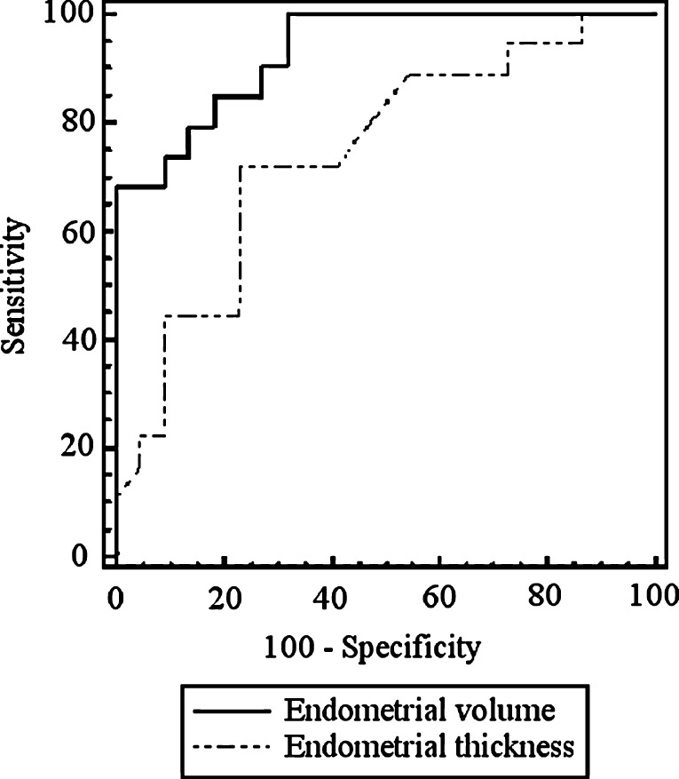
Results: Eighteen patients have achieved pregnancy. A significant difference was found for endometrial volume: 6.49+/-1.97 mL versus 3.40+/-1.11 mL (pregnant versus not pregnant); and thickness: 11.15+/-2.75 mm versus 9.77+/-1.85 mm. The ROC curve was used to detect the best cutoff values: endometrial volume of 3.48 mL (sensitivity-100%, specificity-68.2%) and endometrial thickness of 10.3 mm (sensitivity-72.2%, specificity-77.3%). The area under curve was significant higher for endometrial volume (0.909 versus 0.745, p=0.027). No pregnancy was achieved in women who had an endometrial volume <3.8 mL (15 patients) or thickness <7.9 mm (3 patients).
Conclusions: The endometrial volume and thickness were significant higher in pregnant women and this difference was more prominent for endometrial volume.
Figures
References
-
- Lambers MJ, Van Weering HG, Van't Grunewold MS, Lambalk CB, Homburg R, Schats R, Hompes PG. Optimizing hCG cut-off values: a single determination on day 14 or 15 is sufficient for a reliable prediction of pregnancy outcome. Eur J Obstet Gynecol Reprod Biol 2006 [Epub ahead of print]. - PubMed
-
- Keith SC, London SN, Weitzman GA, O’Brien TJ, Miller MM. Serial transvaginal ultrasound scans and beta-human chorionic gonadotropin levels in early singleton and multiple pregnancies. Fertil Steril. 1993;59:1007–10. - PubMed
MeSH terms
LinkOut - more resources
Full Text Sources
