. 2007 Apr;120(4):573-9.
doi: 10.1111/j.1365-2567.2006.02538.x.

Interleukin-4 inhibition of osteoclast differentiation is stronger than that of interleukin-13 and they are equivalent for induction of osteoprotegerin production from osteoblasts

Affiliations

Interleukin-4 inhibition of osteoclast differentiation is stronger than that of interleukin-13 and they are equivalent for induction of osteoprotegerin production from osteoblasts

Atsushi Yamada et al. Immunology. 2007 Apr.

Abstract

Interleukin (IL)-4 and IL-13 are closely related cytokines known to inhibit osteoclast formation by targeting osteoblasts to produce an inhibitor, osteoprotegerin (OPG), as well as by directly targeting osteoclast precursors. However, whether their inhibitory actions are the same remains unclear. The inhibitory effect of IL-4 was stronger than that of IL-13 in an osteoclast-differentiation culture system containing mouse osteoblasts and osteoclast precursors. Both cytokines induced OPG production by osteoblasts in similar time- and dose-dependent manners. However, IL-4 was stronger in direct inhibition that targeted osteoclast precursors. Furthermore, IL-4 induced phosphorylation of signal transducer and activator of transcription-6 (STAT6) at lower concentrations than those of IL-13 in osteoclast precursors. IL-4 but not IL-13 strongly inhibited the expression of nuclear factor of activated T-cells, cytoplasmic 1 (nuclear factor-ATc1), a key factor of osteoclast differentiation, by those precursors. Thus, the activities of IL-4 and IL-13 toward osteoclast precursors were shown to be different in regards to inhibition of osteoclast differentiation, whereas those toward osteoblasts for inducing OPG expression were equivalent.


Figures

Figure 1
Figure 1
Effects of IL-4 and IL-13 on osteoclast differentiation in a coculture system. (a) BMMs were cocultured with UAMS-32 cells and various concentrations of IL-4 (○) or IL-13 (▵) in the presence of 100 nm 1α,25-(OH)2D3. The same cultures were also performed in the absence of 1α,25-(OH)2D3 (D3) with IL-4 (•) or IL-13 (▴). After culturing the cells, cell lysates were harvested and TRAP activity was measured. Data are shown as the means ± SE (n = 4). (b) BMMs cocultured with primary osteoblasts or UAMS-32 cells in the absence or presence of 10 nm of 1α,25-(OH)2D3 (D3), 10 ng/ml of IL-4 or 10 ng/ml IL-13. The cells were fixed and stained for TRAP (original magnification: ×10).
Figure 2
Figure 2
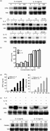
Regulation of OPG and RANKL gene expression by IL-4 and IL-13 in osteoblastic cells. (a) Dose effects of IL-4 and IL-13 on the induction of OPG mRNA. UAMS-32 cells were treated with 1, 10, and 100 ng/ml of IL-4 or IL-13 for 3 hr. (b) Time course analysis of OPG mRNA expression in the presence of IL-4 or IL-13. UAMS-32 cells were treated with 10 ng/ml of IL-4 or IL-13 for 1, 3, 6, 12, and 24 hr. (c) Dose effects of IL-4 and IL-13 on the secretion of OPG protein. UAMS-32 cells were treated with 0·1, 0·5, 1, 10, and 100 ng/ml of IL-4 (white bars) or IL-13 (grey bars) for 12 hr. Data are shown as the means ± SD (n = 3). (d) Time course analysis of the secretion of OPG protein in the presence of IL-4 or IL-13. UAMS-32 cells were treated with 10 ng/ml of IL-4 (black bar) or IL-13 (grey bar) for 6, 12, 24, 36, and 48 hr. Concentrations of OPG in the conditioned media of UAMS-32 cells were determined by enzyme-linked immunosorbent assay. Data are shown as the means ± SD (n = 3). (e) Effects of IL-4 and IL-13 on OPG mRNA in osteoblastic cells pretreated with 1α,25-(OH)2D3. UAMS-32 cells were pretreated with 100 nm of 1α,25-(OH)2D3 for 1 hr, followed by treatment with 10 ng/ml of IL-4 or IL-13 for 3 hr. (f) Effects of IL-4 and IL-13 on RANKL mRNA in osteoblastic cells. UAMS-32 cells were treated with 1, 10, and 100 ng/ml of IL-4 or IL-13 for 24 hr. (a, c, e, and f) The expressions of RANKL mRNA, OPG mRNA, and 18S rRNA were detected by Northern blot analysis.
Figure 3
Figure 3
Effects of IL-4 and IL-13 on osteoclast differentiation induced by RANKL in osteoclast precursor cultures. (a) BMMs were cultured with various concentrations of IL-4 with (○) or without (•) 150 ng/ml of RANKL, or IL-13 with (▵) or without (▴) 150 ng/ml of RANKL. After culturing the cells, the cell lysates were harvested and TRAP activity was measured. Data are shown as the means ± SE (n = 4). (b) Photographs of BMCs and BMMs treated with 10 ng/ml of IL-4 or IL-13. The cells were fixed and stained for TRAP (original magnification ×10).
Figure 4
Figure 4
Expression of receptor components for IL-4 and IL-13 in osteoclast precursors and osteoblastic cells. Total RNAs were extracted from BMCs, primary osteoblasts (OBs), BMMs, RAW264.7 cells, UAMS-32 cells, and ST-2 cells. Subsequently, the mRNA expressions of IL-4Rα, IL-13Rα1, γc, and GAPDH in the cells were analysed by RT–PCR.
Figure 5
Figure 5
Phosphorylation of STAT6 by IL-4 and IL-13 in UAMS-32 cells and BMMs. UAMS-32 (a) cells and BMMs (b) were treated with 0·1, 1, 10 and 10 ng/ml of IL-4 and IL-13 for 30 min. Subsequently, phosphorylation of STAT6 protein was analysed by Western blot analysis.
Figure 6
Figure 6
Effects of IL-4 and IL-13 on NFATc1 mRNA expression in osteoclast precursors. BMMs were treated with or without 150 ng/ml of RANKL in the presence of IL-4 or IL-13 at the indicated concentrations for 24 hr. Total RNA was then extracted and NFATc1 mRNA expression was examined using RT–PCR.

References

    1. Suda T, Takahashi N, Udagawa N, Jimi E, Gillespie MT, Martin TJ. Modulation of osteoclast differentiation and function by the new members of the tumor necrosis factor receptor and ligand families. Endocr Rev. 1999;20:345–57. - PubMed
    1. Simonet WS, Lacey DL, Dunstan CR, et al. Osteoprotegerin: a novel secreted protein involved in the regulation of bone density. Cell. 1997;89:309–19. - PubMed
    1. Tsuda E, Goto M, Mochizuki S, Yano K, Kobayashi F, Morinaga T, Higashio K. Isolation of a novel cytokine from human fibroblasts that specifically inhibits osteoclastogenesis. Biochem Biophys Res Commun. 1997;234:137–42. - PubMed
    1. Yasuda H, Shima N, Nakagawa N, et al. Identity of osteoclastogenesis inhibitory factor (OCIF) and osteoprotegerin (OPG): a mechanism by which OPG/OCIF inhibits osteoclastogenesis in vitro. Endocrinology. 1998;139:1329–37. - PubMed
    1. Bucay N, Sarosi I, Dunstan CR, et al. osteoprotegerin-deficient mice develop early onset osteoporosis and arterial calcification. Genes Dev. 1998;12:1260–8. - PMC - PubMed

Publication types

MeSH terms