Clinical Trial
. 2007 Mar 7:5:13.
doi: 10.1186/1479-5876-5-13.

Calcium sulfate and platelet-rich plasma make a novel osteoinductive biomaterial for bone regeneration

Affiliations
Clinical Trial

Calcium sulfate and platelet-rich plasma make a novel osteoinductive biomaterial for bone regeneration

Giuseppe Intini et al. J Transl Med. .

Abstract

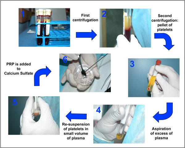
Background: With the present study we introduce a novel and simple biomaterial able to induce regeneration of bone. We theorized that nourishing a bone defect with calcium and with a large amount of activated platelets may initiate a series of biological processes that culminate in bone regeneration. Thus, we engineered CS-Platelet, a biomaterial based on the combination of Calcium Sulfate and Platelet-Rich Plasma in which Calcium Sulfate also acts as an activator of the platelets, therefore avoiding the need to activate the platelets with an agonist.

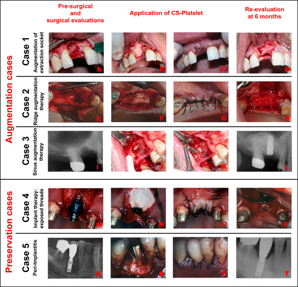
Methods: First, we tested CS-Platelet in heterotopic (muscle) and orthotopic (bone) bone regeneration bioassays. We then utilized CS-Platelet in a variety of dental and craniofacial clinical cases, where regeneration of bone was needed.

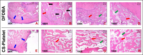
Results: The heterotopic bioassay showed formation of bone within the muscular tissue at the site of the implantation of CS-Platelet. Results of a quantitative orthotopic bioassay based on the rat calvaria critical size defect showed that only CS-Platelet and recombinant human BMP2 were able to induce a significant regeneration of bone. A non-human primate orthotopic bioassay also showed that CS-Platelet is completely resorbable. In all human clinical cases where CS-Platelet was used, a complete bone repair was achieved.

Conclusion: This study showed that CS-Platelet is a novel biomaterial able to induce formation of bone in heterotopic and orthotopic sites, in orthotopic critical size bone defects, and in various clinical situations. The discovery of CS-Platelet may represent a cost-effective breakthrough in bone regenerative therapy and an alternative or an adjuvant to the current treatments.


Figures

Figure 1
Figure 1
Preparation of CS-Platelet: Step 1: aliquots of whole blood (as indicated in Table 1) were collected in tubes containing acid-citrate-dextrose as an anti-coagulant. Step 2: immediately after being drawn, blood was centrifuged (see Table 1) to separate RBCs from platelets and plasma. Step 3: The supernatant composed of platelets and plasma was collected and centrifuged again (see Table 1) in order to pellet the platelets. Step 4 and 5: After this second centrifugation, the platelets were re-suspended in an appropriate volume of autologous plasma to achieve a platelet concentration 8–10 fold above the physiologic levels. (Step 6): CS (powder) was mixed with PRP (liquid) in a ratio of1 g of CS to 240 μl of PRP.
Figure 2
Figure 2
Heterotopic (muscle) bioassay in ferrets: (A): The biceps femoris of a ferret is shown. A sample of CS-Platelet and a sample of CS were implanted in the muscle tissue. (B): Four weeks after implantation the x-ray evaluations showed the presence of radio-opaque formations only in correlation with the implantations of CS-Platelet. (C, E-H): At the histological evaluation, the radio-opaque formation appeared as an ossicle formed by trabecular bone. (D, I-L): No bone formation was observed in correspondence with the implantations of Calcium Sulfate alone. Hematoxylin and Eosin staining.
Figure 3
Figure 3
Orthotopic (bone) bioassay in rats – From left to right: Microcomputed Tomography (μCT) images, sagittal histological sections, higher magnifications of histological sections (100× and 400× original magnifications). Eight weeks after implantation in the 8 mm rat critical size defects, only CS-Platelet and rhBMP2 showed a regeneration of bone extended at the center of the defects. In all other groups a limited regeneration of bone was seen only at the periphery of the defects. Goldner's trichrome staining: nuclear chromatin (brown-black), cytoplasm (bright red), erythrocytes (orange), collagen (light green), mineralized bone (green), osteoid (red).
Figure 4
Figure 4
Orthotopic (bone) bioassay in rats – Quantitative analysis of Tomography Data. Volume (A): only CS-Platelet, rhBMP2, and the External control showed a statistically significant difference from the negative control (p < 0.001). Surface area (B): only CS-Platelet, rhBMP2, and the External control showed a statistically significant difference from the negative control (p < 0.001). Density (C): all treatment groups showed a density of the regenerated bone statistically different from the density of the bone normally present in an area of 8 mm of the calvaria (External control)(p < 0.001).
Figure 5
Figure 5
Non-human primate orthotopic (bone) bioassay – Histology. (A-D): samples of alveolar bone implanted with DFDBA. (E-H): Samples of alveolar bone implanted with CS-Platelet. All grafted sites, whether implanted with CS-Platelet or with DFDBA, showed regeneration of trabecular bone. In the sites grafted with DFDBA some residual biomaterial was still visible (B, black arrows). Hematoxylin and Eosin staining. (A and E, 40× original magnification). (B-D and F-H, 100× original magnification).
Figure 6
Figure 6
Human clinical cases (from the top to the bottom). Augmentation cases: (A-D): Augmentation of extraction socket upon extraction of tooth # 8. (E-H): Ridge augmentation therapy in area of teeth # 5–6. (I-L): Sinus augmentation therapy (induced bone formation within the right maxillary sinus) prior insertion of a titanium implant in area of teeth # 3–4. Preservation cases: (M-P): Regeneration of bone around the exposed threads of a titanium implant positioned in area of tooth # 9. (Q-T): Regeneration of bone around a titanium implant in position of tooth # 30 affected by peri-implantitis.

References

    1. Weinstein SL. 2000-2010: the bone and joint decade. J Bone Joint Surg Am. 2000;82:1–3. doi: 10.1302/0301-620X.82B1.10822. - DOI - PubMed
    1. Katz SI. Boning up for health: the national bone and joint decade. J Bone Miner Res. 2004;19:1605–1606. doi: 10.1359/JBMR.040810. - DOI - PubMed
    1. Bush GW. National bone and joint decade: 2002-2011. A proclamation by the President of the United States of America. J Bone Joint Surg Am. 2002;84-A:1297; discussion 1298. - PubMed
    1. Urist MR. Bone: formation by autoinduction. Science. 1965;150:893–899. doi: 10.1126/science.150.3698.893. - DOI - PubMed
    1. Urist MR, Huo YK, Brownell AG, Hohl WM, Buyske J, Lietze A, Tempst P, Hunkapiller M, DeLange RJ. Purification of bovine bone morphogenetic protein by hydroxyapatite chromatography. Proc Natl Acad Sci U S A. 1984;81:371–375. doi: 10.1073/pnas.81.2.371. - DOI - PMC - PubMed

Publication types

LinkOut - more resources