. 2007 May;81(10):5315-24.
doi: 10.1128/JVI.02129-06. Epub 2007 Mar 7.

Influenza virus infection causes specific degradation of the largest subunit of cellular RNA polymerase II

Affiliations

Influenza virus infection causes specific degradation of the largest subunit of cellular RNA polymerase II

A Rodriguez et al. J Virol. 2007 May.

Erratum in

  • J Virol. 2008 Apr;82(7):3811

Abstract

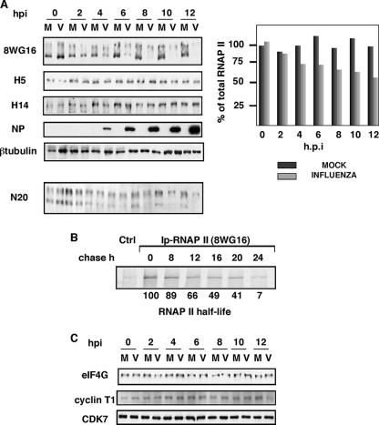
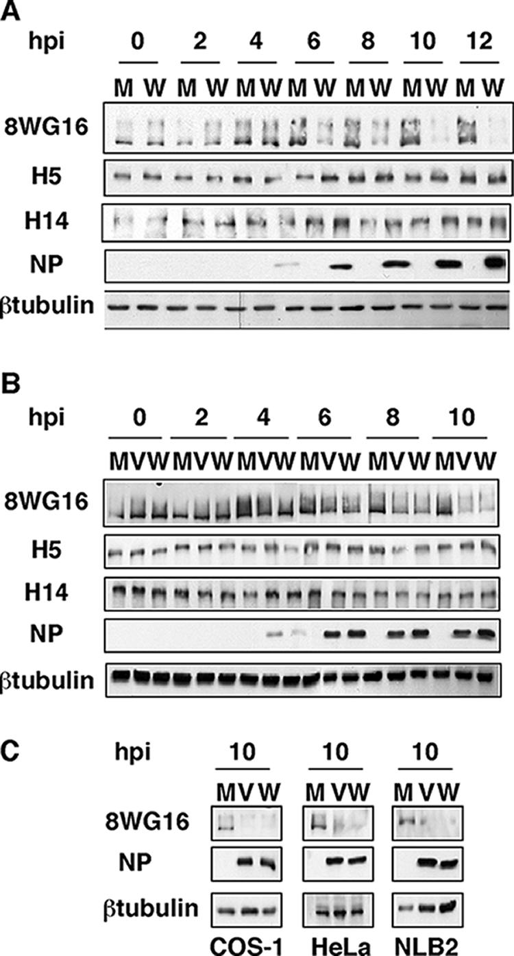
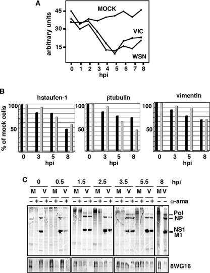
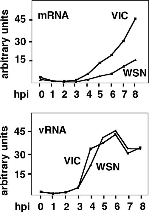
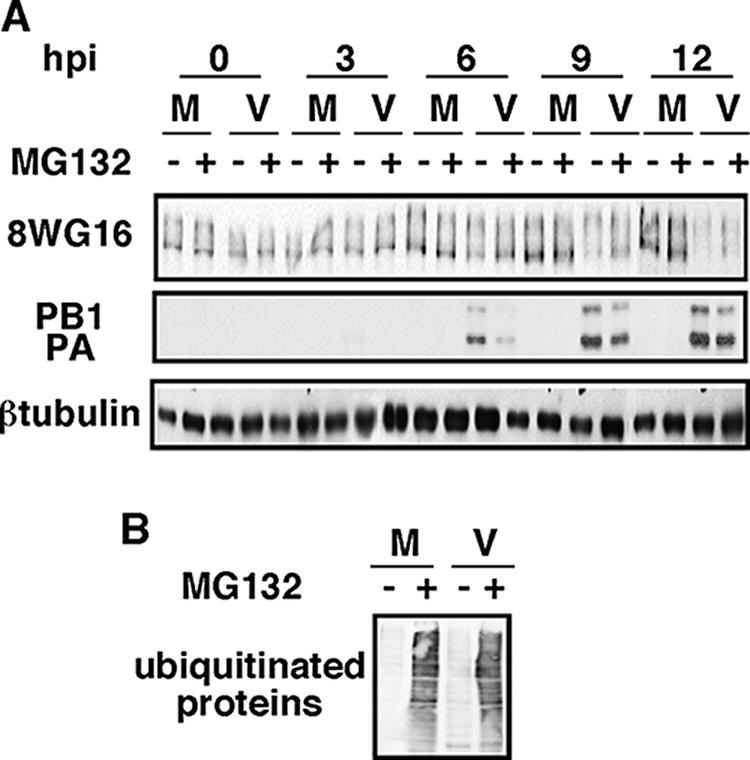
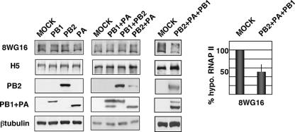
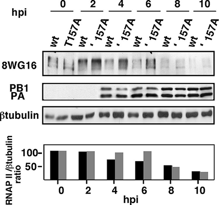
It has been described that influenza virus polymerase associates with RNA polymerase II (RNAP II). To gain information about the role of this interaction, we explored if changes in RNAP II occur during infection. Here we show that influenza virus causes the specific degradation of the hypophosphorylated form of the largest subunit of RNAP II without affecting the accumulation of its hyperphosphorylated forms. This effect is independent of the viral strain and the origin of the cells used. Analysis of synthesized mRNAs in isolated nuclei of infected cells indicated that transcription decreases concomitantly with RNAP II degradation. Moreover, this degradation correlated with the onset of viral transcription and replication. The ubiquitin-mediated proteasome pathway is not involved in virally induced RNAP II proteolysis. The expression of viral polymerase from its cloned cDNAs was sufficient to cause the degradation. Since the PA polymerase subunit has proteolytic activity, we tested its participation in the process. A recombinant virus that encodes a PA point mutant with decreased proteolytic activity and that has defects in replication delayed the effect, suggesting that PA's contribution to RNAP II degradation occurs during infection.


Figures

FIG. 1.
FIG. 1.
Influenza virus infection produces degradation of hypophosphorylated RNAP II. (A) HEK293T cells were infected with the VIC strain of influenza virus and at the designated hpi hypophosphorylated RNAP II (8WG16), RNAP II Ser-2P and Ser-5P (H5), RNAP II Ser-5P (H14), total RNAP II (N20), and the indicated proteins were monitored in total cell extracts by Western blotting. Five different experiments were carried out, and a representative experiment is shown. Quantitation of the amount of total RNAP II (hypo- plus hyperphosphoryalated forms) during the virus infection is shown at the right. M, mock-infected cells; V, influenza virus-infected cells. (B) Pulse-chase experiments. Synthesis of hypophosphorylated RNAP II was monitored in HEK293T cells with a mixture of [35S]Met/Cys. The specific 8WG16 antibody was used to immunoprecipitate (Ip) hypophosphorylated RNAP II. Ctrl, unspecific monoclonal antibody. (C) HEK293T cells were infected with the VIC strain of influenza virus and at the designated hpi the presence of the indicated proteins was monitored in total cell extracts by Western blotting.
FIG. 2.
FIG. 2.
Degradation of hypophosphorylated RNAP II in different virus-host cell systems. (A) HEK293T cells were mock infected (M) or infected with the WSN strain of influenza virus (W) and processed as for Fig. 1. (B) NIH 3T3 cells were mock infected or infected with the VIC (V) or the WSN strain of influenza virus and processed as for Fig. 1. (C) COS-1, HeLa, and NLB2 cells were mock infected or infected with the VIC or WSN strain of influenza virus, and at 10 hpi the presence of RNAP II (8WG16) and the designated proteins was analyzed by Western blotting.
FIG. 3.
FIG. 3.
Dependence of cellular mRNA and viral protein synthesis on active RNAP II. (A) Synthesis of cellular mRNA. HEK293T cells were mock-infected or infected with the VIC or WSN strain of influenza virus, and at the indicated hpi nuclei were isolated and total RNA synthesis was measured by in vitro incorporation of a labeled ribonucleotide with or without α-amanitin (5 μg/ml). The mRNA synthesis due to RNAP II activity is calculated as the difference between the values obtained without the drug (total synthesis is synthesis of RNAP I plus RNAP II plus RNAP III) and with the drug (synthesis of RNAP I plus RNAP III). In the infected cells the contribution of viral replication that is insensitive to α-amanitin should be taken into account. (B) Synthesis of hStaufen-1, β-tubulin, and vimentin mRNAs. Total labeled RNAs from panel A were used as probes for runoff experiments. Five hundred nanograms of the corresponding coding sequences was used, and the label was quantitated in a phosphorimager. Black bars, cells infected with the VIC strain; gray bars, cells infected with the WSN strain. (C) HEK293T cells were mock infected (M) or infected with the VIC strain of influenza virus (V), and α-amanitin (50 μg/ml; α-ama) was added (+) or not (−) to the cell culture at the indicated hpi. At 7 hpi the cells were metabolically labeled with [35S]Met-Cys. Cells were collected at 8 hpi and the synthesized proteins analyzed. At the same times, total cell extracts were used to analyze hypophosphorylated RNAP II by Western blotting (8WG16).
FIG. 4.
FIG. 4.
Degradation of hypophosphorylated RNAP II correlates with the onset of viral transcription and replication. HEK293T cells were mock infected or infected with the VIC or WSN strain of influenza virus, and at the indicated hpi total RNAs were isolated and used to detect the positive sense RNA (top) or the vRNA (vRNA; bottom) of viral nucleoproteins by Northern blot analysis.
FIG. 5.
FIG. 5.
The proteasome pathway is not involved in the degradation of hypophosphorylated RNAP II. (A) HEK293T cells were mock infected (M) or infected with the VIC strain (V), with (+) or without (−) MG132, and at the indicated hpi hypophosphorylated RNAP II (8WG16) and the designated proteins were detected by Western blotting. (B) Western blot using antiubiquitin antibody of mock-infected cells and cells infected at 10 hpi from panel A.
FIG. 6.
FIG. 6.
Reconstituted viral polymerase degrades hypophosphorylated RNAP II. HEK293T cells were transfected with plasmids expressing PA, PB1, or PB2 individually (left), with combinations of two of them (middle), or with plasmids expressing PA, PB1, and PB2 together (right), and at 16 h posttransfection the amounts of hypophosphorylated RNAP II (8WG16), hyperphosphorylated RNAP II (H5), and the indicated proteins were detected by Western blotting. Quantitation of RNAP II degradation under conditions of polymerase reconstitution is shown at the right (means and standard deviations).
FIG. 7.
FIG. 7.
The PA subunit is involved in hypophosphorylated RNAP II degradation. HEK293T cells were infected with rescued recombinant viruses containing wild-type PA (wt) or T157A mutated PA (T157A), and at different hpi hypophosphorylated RNAP II (8WG16) and the indicated proteins were detected by Western blot assays. (Bottom) Quantitation of hypophosphorylated RNAP II levels during the infection. The ratios of RNAP II/β-tubulin accumulation are shown. Black bars, wt PA; gray bars, PA T157A. Three different experiments were carried out, and a representative experiment is shown.

References

    1. Aragón, T., S. de la Luna, I. Novoa, L. Carrasco, J. Ortín, and A. Nieto. 2000. Eukaryotic translation initiation factor 4GI is a cellular target for NS1 protein, a translational activator of influenza virus. Mol. Cell. Biol. 20:6259-6268. - PMC - PubMed
    1. Arrese, M., and A. Portela. 1996. Serine 3 is critical for phosphorylation at the N-terminal end of the nucleoprotein of influenza virus A/Victoria/3/75. J. Virol. 70:3385-3391. - PMC - PubMed
    1. Banerjee, R., M. K. Weidman, A. Echeverri, P. Kundu, and A. Dasgupta. 2004. Regulation of poliovirus 3C protease by the 2C polypeptide. J. Virol. 78:9243-9256. - PMC - PubMed
    1. Bárcena, J., D. l. L. S., M. Ochoa, J. A. Melero, A. Nieto, J. Ortín, and A. Portela. 1994. Monoclonal antibodies against the influenza virus PB2 and NP polypeptides interfere with the initiation step of viral mRNA synthesis in vitro. J. Virol. 68:6900-6909. - PMC - PubMed
    1. Bark-Jones, S. J., H. M. Webb, and M. J. West. 2006. EBV EBNA 2 stimulates CDK9-dependent transcription and RNA polymerase II phosphorylation on serine 5. Oncogene 25:1775-1785. - PubMed

Publication types

MeSH terms

LinkOut - more resources