. 2007 May;81(10):5155-65.
doi: 10.1128/JVI.01796-06. Epub 2007 Mar 7.

Incorporation of human immunodeficiency virus type 1 reverse transcriptase into virus-like particles

Affiliations

Incorporation of human immunodeficiency virus type 1 reverse transcriptase into virus-like particles

Wei-Hao Liao et al. J Virol. 2007 May.

Abstract

We demonstrate that a genetically engineered human immunodeficiency virus type 1 (HIV-1) reverse transcriptase (RT) composed mainly of p66 or p51 subunits can be incorporated into virus-like particles (VLPs) when coexpressed with HIV-1 Pr55(gag). VLP-associated RT exhibited a detergent-resistant association with immature cores during sucrose gradient equilibrium centrifugation, suggesting that RT is incorporated into VLPs. However, RT that retains downstream integrase (IN) is severely inhibited in terms of incorporation into VLPs. Results from immunofluorescence tests reveal that RT-IN is primarily localized at the perinuclear area and exhibits poor colocalization with Gag. IN removal leads to a redistribution of RT throughout the cytoplasm and improved RT incorporation into VLPs. Similar results were observed for RT-IN in which alanine was substituted for 186-Lys-Arg-Lys-188 residues of the IN putative nuclear localization signal, suggesting that IN karyophilic properties may partly account for the inhibitory effect of IN on RT incorporation. Although the membrane-binding capacity of RT was markedly reduced compared to that of wild-type Gag or Gag-Pol, the correlation of membrane-binding ability with particle incorporation efficiency was incomplete. Furthermore, we observed that membrane-binding-defective myristylation-minus RT can be packaged into VLPs at the same level as its normal myristylated counterpart. This suggests that the incorporation of RT into VLPs is independent of membrane affinity but very dependent on RT-Gag interaction. Results from a genetic analysis suggest that the Gag-interacting regions of RT mainly reside in the thumb subdomain and that the RT-binding domains of Gag are located in the matrix (MA) and p6 regions.


Figures

FIG. 1.
FIG. 1.
Schematic representatives of HIV-1 Gag, Gag-Pol, and RT-IN expression constructs. pGAG and GPfs express Pr55gag and Pr160gag-pol, respectively. Indicated are the HIV Gag protein domains MA (matrix), CA (capsid), NC (nucleocapsid), and p6 and the pol-encoded p6*, PR, RT, and IN; numbers refer to amino acid residue positions. p66RT subdomain boundaries are also indicated. Dashed lines represent deleted sequences. Designated construct numbers indicate IN or RT terminal amino acid residue positions. The final three or four amino acid residues in each truncated construct are shown; inserted or altered amino acids are in boldface.
FIG. 2.
FIG. 2.
HIV-1 RT expression and incorporation into VLPs. 293T cells were cotransfected with 10 μg pGAG and 10 μg of PR-defective GPfs, GPfsPR, or the indicated construct. Cells and supernatant were collected at 48 h posttransfection for protein analysis as described in Materials and Methods. Cell samples corresponding to 4% of total cell lysates and supernatant samples corresponding to 50% of total recovered viral pellets were fractionated by 10% SDS-PAGE. (A) HIV-1 RT and Gag-Pol were probed with an anti-RT polyclonal antibody. Since R182 is smaller in size, we must consider the possibility that it was electrophoresed off the gel (lane 17). However, even when we repeated this experiment and carefully monitored the gel, we still failed to detect R182. (B) HA-tagged RT proteins were detected with an anti-HA monoclonal antibody. Membranes from top panels A and B were stripped and reprobed using a monoclonal antibody directed against HIV-1 p24CA. Note that the Pr160gag-pol bands (B, lanes 1 and 10) did not appear when probed with the anti-HA antibody but were detected by the anti-p24CA antibody. Molecular size marker positions are shown on the right.
FIG. 3.
FIG. 3.
Sucrose density gradient fractionation of virus-associated HIV-1 RT. 293T cells were cotransfected with HA-tagged R560 (hR560) and pGAG. At 48 h posttransfection, culture supernatant was collected, filtered, and pelleted through 20% sucrose cushions. Viral pellets were resuspended and centrifuged through 30 to 70% sucrose gradients containing a layer of 1% Triton X-100 as described in Materials and Methods. Ten fractions of equal size were collected from the top of each gradient after 16 h of centrifugation. In addition to measuring the density of each fraction, we used Western immunoblotting to analyze Gag and RT protein levels. Fraction densities (in g/ml) are indicated at the top.
FIG. 4.
FIG. 4.
Membrane flotation analysis of RT proteins. 293T cells were transfected with pGAG, GPfsPR, or the designated expression constructs. Two days posttransfection, cells were harvested, homogenized, and subjected to equilibrium flotation centrifugation analysis as described in Materials and Methods. Ten fractions were collected from the top downward. Fraction aliquots were resolved by SDS-PAGE (10%) and probed with a monoclonal antibody directed against HIV-1 p24CA or the HA tag. During ultracentrifugation, membrane-bound Gag proteins floated to the 10 to 65% sucrose interface and became enriched in fraction 3.
FIG. 5.
FIG. 5.
Subcellular localization of HIV-1 RT. HeLa cells grown on coverslips were transfected with pGAG (A), PR-defective GPfs (B), hRN288 (C), hRN198 (D), hR560 (E), hR515 (F), hR425 (G) hR305 (H), or hR182 (I) or transfected with pGAG plus hRN288 (J), hRN198 (M), or hR560 (P). At 48 h posttransfection, cells were fixed and permeabilized for immunofluorescent staining as described in Materials and Methods. Primary antibodies were either mouse anti-HIV-1 p24CA (A and B) or anti-HA (C to I); the secondary antibody was rhodamine-conjugated rabbit anti-mouse. For double staining (J to R), HIV-1 Gag was detected with the anti-HIV-1 p24CA monoclonal antibody followed by the rhodamine-conjugated rabbit anti-mouse antibody (J, M, and P); the HA-tagged proteins were probed with a fluorescein isothiocyanate-conjugated anti-HA antibody (K, N, and Q). Merged green and red fluorescence images are shown in panels L, O, and R. Mock-transfected HeLa cells and cells not exposed to the primary antibody yielded no signal (data not shown). Bar, 10 μm.
FIG. 6.
FIG. 6.
Effect of IN putative nuclear localization mutation on RT-IN subcellular localization and particle incorporation. (A and B) Subcellular localization of HIV-1 RT-IN. HeLa cells were transfected with hRN288 (A) or hRN(NLS) (B); the two are identical except for alanine substitutions for the 186-Lys-Arg-Lys-188 residues in the IN nuclear localization motif. HA-tagged RT-IN proteins were detected as described in the legend for Fig. 5. Bar, 10 μm. (C) Incorporation of HIV-1 RT-IN into VLPs. 293T cells were cotransfected with pGAG and each indicated construct. At 48 h posttransfection, cells and culture supernatant were collected, prepared, and subjected to Western immunoblotting. HA-tagged RT proteins and HIV-1 Gag were probed with anti-HA and anti-p24CA antibodies, respectively.
FIG. 7.
FIG. 7.
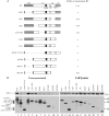
Mapping of RT-binding domains on Gag. (A) Schematic representatives of HIV-1 gag mutants. Four major domains are indicated: MA, CA, NC, and p6. Also indicated are the space peptides SP1 and SP2 and a major homology region (MHR) in CA. Each Gag mutant contains an internal deletion (dashed line) and/or a C-terminal truncation mutation in the gag coding sequence. The ability of each mutant to incorporate HIV-1 RT is summarized on the right as follows: +++, RT incorporation efficiency comparable to that of wt (≥80% of wt); +, efficiency between 2 and 10% of wt; NA, not applicable. Note that MT380 is severely defective in VLP production. (B) Incorporation of HIV-1 RT into virus-like particles. 293T cells were cotransfected with hR560 and the designated constructs. Two days posttransfection, culture supernatant and cells were collected, prepared, and subjected to 10% SDS-PAGE. hR560 and Gag proteins were probed with anti-HA and anti-p24CA monoclonal antibodies, respectively. Levels of virus-associated Gag and RT in each sample were quantified by scanning immunoblot band densities. RT/Gag protein level ratios were calculated for each sample and normalized to that of wt in parallel experiments. Percentages of virus-associated RT denote the ability of each mutant to package RT. Molecular mass marker positions are shown on the right. Predicted molecular masses corresponding to Gag deletion mutant sizes were as follows: Δ241-282, 51 kDa; ΔNP and ΔNC, 50 kDa; T449, 49 kDa; T431, 47 kDa; ΔMA, 44 kDa; MT449, 39 kDa; MT431, 37 kDa; Δ(MA+2/3CA) and MT380, 27 to 28 kDa. Note that the immunoblot shown in panel B (left) was intentionally overexposed to allow the assembly-defective gag mutant to be viewed. p24-associated degraded bands usually appeared as exposure was increased.
FIG. 8.
FIG. 8.
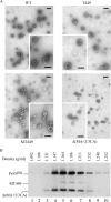
Analysis of HIV-1 Gag VLPs. (A) Transmission electron microscopy of concentrated culture supernatant from 293T cells expressing the wt or the indicated gag deletion mutant. Bars, 200 nm. (B) Sucrose density gradient fractionation of HIV-1 Gag VLPs. 293T cells were transfected with wt, MT449, or Δ(MA+2/3CA). At 48 to 72 h posttransfection, culture supernatants were collected and pelleted through 20% sucrose cushions. Viral pellets were suspended in TSE buffer. To make direct comparisons with wt HIV-1 particle densities, wt viral pellets were spun through the same sucrose density gradient (20 to 60%) with pooled pellets containing MT449 and Δ(MA+2/3CA) at 274,000 × g for 16 h. Fractions were collected, measured for sucrose density, and analyzed for Gag protein levels by immunoblotting. Fraction densities are indicated at the top. The relatively lower signal of MT449 was due to loss of the viral pellets prior to pooling them together.

Similar articles

Cited by

References

    1. Abdurahman, S., S. Hoglund, L. Goobar-Larsson, and A. Vahlne. 2004. Selected amino acid substitutions in the C-terminal region of human immunodeficiency virus type 1 capsid protein affect virus assembly and release. J. Gen. Virol. 85:2903-2913. - PubMed
    1. Accola, M. A., S. Hoglund, and H. G. Gottlinger. 1998. A putative α-helical structure which overlaps the capsid-p2 boundary in the human immunodeficiency virus type 1 Gag precursor is critical for viral particle assembly. J. Virol. 72:2072-2078. - PMC - PubMed
    1. Accola, M. A., B. Strack, and H. G. Gottlinger. 2000. Efficient particle production by minimal Gag constructs which retain the carboxy-terminal domain of human immunodeficiency virus type 1 capsid-p2 and a late assembly domain. J. Virol. 74:5395-5402. - PMC - PubMed
    1. Borsetti, A., A. Ohagen, and H. G. Gottlinger. 1998. The C-terminal half of the human immunodeficiency virus type 1 Gag precursor is sufficient for efficient particle assembly. J. Virol. 72:9313-9317. - PMC - PubMed
    1. Bryant, M., and L. Ratner. 1990. Myristoylation-dependent replication and assembly of human immunodeficiency virus 1. Proc. Natl. Acad. Sci. USA 87:523-527. - PMC - PubMed

Publication types

MeSH terms

Substances