

Neurovascular coupling is not mediated by potassium siphoning from glial cells
- PMID: 17344384
- PMCID: PMC2289782
- DOI: 10.1523/jneurosci.3204-06.2007
Neurovascular coupling is not mediated by potassium siphoning from glial cells
Abstract
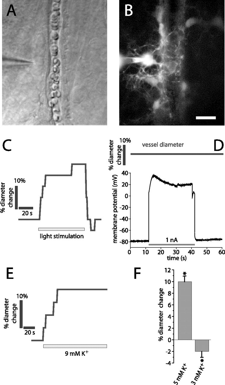
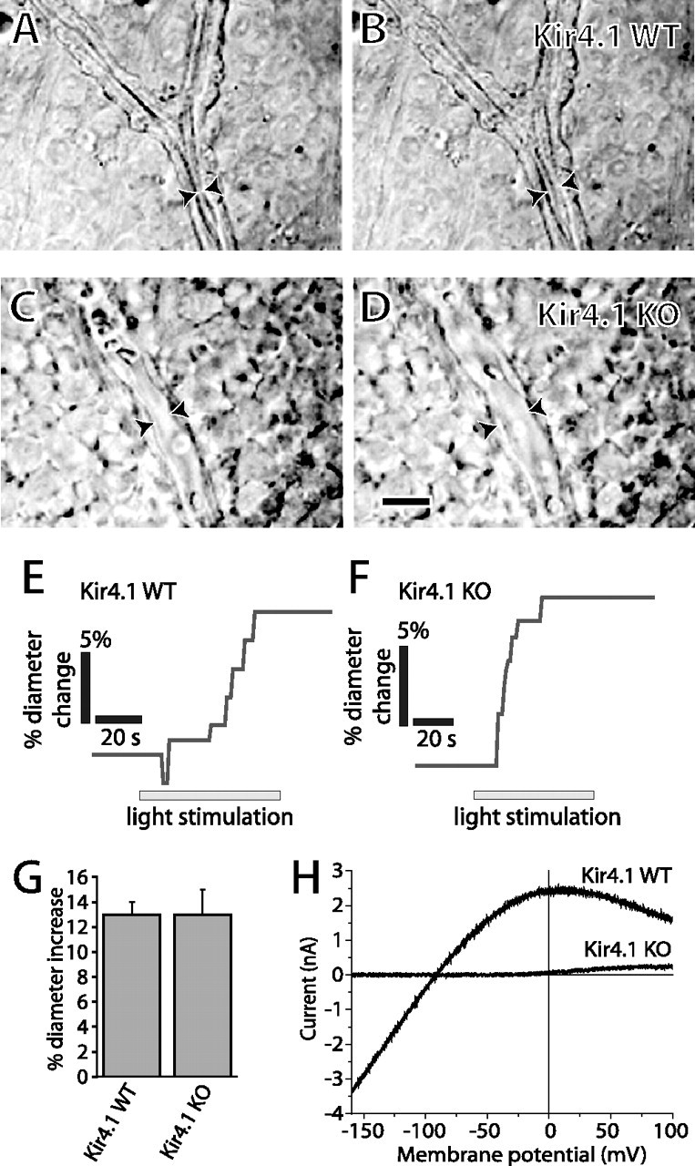
Neuronal activity evokes localized changes in blood flow, a response termed neurovascular coupling. One widely recognized hypothesis of neurovascular coupling holds that glial cell depolarization evoked by neuronal activity leads to the release of K+ onto blood vessels (K+ siphoning) and to vessel relaxation. We now present two direct tests of this glial cell-K+ siphoning hypothesis of neurovascular coupling. Potassium efflux was evoked from glial cells in the rat retina by applying depolarizing current pulses to individual cells. Glial depolarizations as large as 100 mV produced no change in the diameter of adjacent arterioles. We also monitored light-evoked vascular responses in Kir4.1 knock-out mice, where functional Kir K+ channels are absent from retinal glial cells. The magnitude of light-evoked vasodilations was identical in Kir4.1 knock-out and wild-type animals. Contrary to the hypothesis, the results demonstrate that glial K+ siphoning in the retina does not contribute significantly to neurovascular coupling.
Figures
References
-
- Brew H, Gray PTA, Mobbs P, Attwell D. Endfeet of retinal glial cells have higher densities of ion channels that mediate K+ buffering. Nature. 1986;324:466–468. - PubMed
-
- Bringmann A, Faude F, Reichenbach A. Mammalian retinal glial (Müller) cells express large-conductance Ca2+-activated K+ channels that are modulated by Mg2+ and pH and activated by protein kinase A. Glia. 1997;19:311–323. - PubMed
-
- Edvinsson L, MacKenzie ET, McCulloch J. Cerebral blood flow and metabolism. New York: Raven; 1993.
Publication types
MeSH terms
Substances
Grants and funding
LinkOut - more resources
Full Text Sources
Medical
Molecular Biology Databases