. 2007 Mar 7;27(10):2472-82.
doi: 10.1523/JNEUROSCI.2279-06.2007.

Impaired cerebellar development and function in mice lacking CAPS2, a protein involved in neurotrophin release

Affiliations

Impaired cerebellar development and function in mice lacking CAPS2, a protein involved in neurotrophin release

Tetsushi Sadakata et al. J Neurosci. .

Abstract

Ca2+-dependent activator protein for secretion 2 (CAPS2/CADPS2) is a secretory granule-associated protein that is abundant at the parallel fiber terminals of granule cells in the mouse cerebellum and is involved in the release of neurotrophin-3 (NT-3) and brain-derived neurotrophic factor (BDNF), both of which are required for cerebellar development. The human homolog gene on chromosome 7 is located within susceptibility locus 1 of autism, a disease characterized by several cerebellar morphological abnormalities. Here we report that CAPS2 knock-out mice are deficient in the release of NT-3 and BDNF, and they consequently exhibit suppressed phosphorylation of Trk receptors in the cerebellum; these mice exhibit pronounced impairments in cerebellar development and functions, including neuronal survival, differentiation and migration of postmitotic granule cells, dendritogenesis of Purkinje cells, lobulation between lobules VI and VII, structure and vesicular distribution of parallel fiber-Purkinje cell synapses, paired-pulse facilitation at parallel fiber-Purkinje cell synapses, rotarod motor coordination, and eye movement plasticity in optokinetic training. Increased granule cell death of the external granular layer was noted in lobules VI-VII and IX, in which high BDNF and NT-3 levels are specifically localized during cerebellar development. Therefore, the deficiency of CAPS2 indicates that CAPS2-mediated neurotrophin release is indispensable for normal cerebellar development and functions, including neuronal differentiation and survival, morphogenesis, synaptic function, and motor learning/control. The possible involvement of the CAPS2 gene in the cerebellar deficits of autistic patients is discussed.


Figures
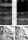
Figure 1.
Figure 1.
Distribution of CAPS family proteins and aberrant lobulation in the CAPS2−/− mouse cerebellum. A, B, To generate CAPS2 knock-out mice, the SmaI–SmaI fragment containing full-length CAPS2 exon 1 was replaced by the Pgk–neo gene cassette (see Materials and Methods). Sagittal sections of P8 wild-type (A) and CAPS2−/− (B) cerebella were immunolabeled with an anti-CAPS2 antibody. Confocal fluorescence images were taken from the cerebellar cortex. C, D, Sagittal sections of P8 wild-type (C) and CAPS2−/− (D) cerebella were immunolabeled with an anti-CAPS1 antibody. PCL, Purkinje cell layer. Scale bars, 20 μm. E, F, Sagittal sections of P28 wild-type (E) and CAPS2−/− (F) cerebella were stained with cresyl violet. Arrows point to the intercrural fissure that normally separates lobules VI and VII. Scale bars, 250 μm.
Figure 2.
Figure 2.
Impaired dendritic arborization of Purkinje cells and delayed granule cell migration in CAPS2−/− mouse cerebellum. A, B, Sagittal sections of P8 wild-type (A) and CAPS2−/− (B) cerebella were immunolabeled with an anti-calbindin antibody. Fluorescence images near the bottom of the primary fissure. Scale bars, 50 μm. C, D, Dendritic arbors of P8 wild-type (C) and CAPS2−/− (D) Purkinje cells in lobule V were visualized by Golgi staining. Scale bars, 10 μm. E, F, Sagittal sections of P17 wild-type (E) and CAPS2−/− (F) cerebella were stained with cresyl violet. Images of the cerebellar cortex layer in lobule VI. The EGL is indicated by a square bracket. Scale bars, 25 μm. G, Statistical analysis of Purkinje cell dendrites. The length of the dendrites was measured at four sites of primary fissure on four sections prepared from different animals (P8). H, Statistical analysis of EGL thickness. The thickness of the EGL was measured at three sites of lobule VI on three sections prepared from different animals (P17). The error bars indicate the SD. **p < 0.01 by Student's t test.
Figure 3.
Figure 3.
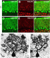
Impaired dendritic arborization of Purkinje cells in the cerebella of P28 CAPS2−/− mice. A–F, Sagittal sections of P28 wild-type (A–C) and CAPS2−/− (D–F) mouse cerebella were immunolabeled with an anti-calbindin antibody (green) and an anti-VGLUT2 (red) antibody. Images of lobule VIII of the cerebellar cortex. Scale bars, 20 μm. G, H, The dendritic arbors of P28 wild-type (G) and CAPS2−/− (H) Purkinje cells were visualized by Golgi staining. Scale bars, 10 μm.
Figure 4.
Figure 4.
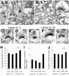
Aberrantly distributed and fewer vesicles within enlarged PF terminals (P15) and an increased number of divided PSDs (P84) in the CAPS2−/− mouse cerebellum. A, B, Electron micrographs of a PF–PC synapse in the P15 wild-type cerebellum (A) and another in the P15 CAPS2−/− cerebellum (B). The insets show higher magnifications. Asterisks represent presynaptic terminals. Scale bars: 500 nm; insets, 200 nm. C–G, Electron micrographs of a PF–PC synapse in the P84 wild-type cerebellum (C, E) and another in the P84 CAPS2−/− cerebellum (D, F, G). Images of lobules IV–V (C, D) and lobules VI–VII (E–G) of the cerebellar cortex. Arrows indicate the positions of perforations. Scale bars: 250 nm. H, Diameter of presynapses. The error bars indicate the SEM. **p < 0.01 by Student's t test. I, The ratio of spines containing divided PSDs to the total spines examined. Multiple PSDs in one synapse is defined as a divided PSD. J, Number of PSDs per spine. The error bars indicate the SEM. **p < 0.01 by F test. Number of synapses examined (n) is indicated.
Figure 5.
Figure 5.
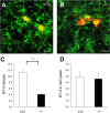
Decreased immunoreactivity of internalized NT-3 in the Purkinje cells of a CAPS2−/− primary culture (confocal image) and decreased NT-3 release activity in the culture. A, B, Cerebellar primary cultures (DIV8) from wild-type mice (A) and CAPS2−/− mice (B) were immunolabeled with an anti-NT-3 antibody (green) and an anti-calbindin (red) antibody. Scale bars, 20 μm. C, NT-3 release activity in the wild-type (open bars) and CAPS2−/− (filled bars) cerebellum was evaluated by measuring the levels of NT-3 spontaneously secreted into the culture medium by cerebellar primary dissociation cultures at DIV8 with an enzyme immunoassay. The y-axis shows NT-3 density corrected for whole-cell density (arbitrary unit). D, The amount of NT-3 in the cell lysates of wild-type (open bars) and CAPS2−/− (filled bars) cultures was evaluated as indicated in C. The y-axis shows NT-3 density corrected for whole-cell density (arbitrary unit). The error bars indicate the SD. **p < 0.01 by Student's t test.
Figure 6.
Figure 6.
Decreased immunoreactivity of phosphorylated Trk (Tyr490) in the Purkinje cell dendrites of CAPS2−/− mice. A–F, Sagittal sections of P8 wild-type (A–C) and CAPS2−/− (D–F) cerebella were immunolabeled with an anti-phosphorylated Trk antibody (green) and an anti-calbindin (red) antibody. Images of lobule IX of the cerebellar cortex are shown. Similar staining patterns were observed throughout the cerebellar cortices of wild-type and CAPS2−/− mice. P, Purkinje cell layer. Scale bars, 25 μm.
Figure 7.
Figure 7.
Apoptotic cell density in the CAPS2−/− mouse cerebellum. A, B, Apoptotic cell density in the EGL (A) and IGL (B) of each lobule of the wild-type (open bars) and CAPS2−/− (filled bars) cerebellum (TUNEL assay). The error bars indicate the SD. **p < 0.01 by Student's t test.
Figure 8.
Figure 8.
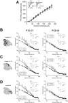
Impairment of short-term synaptic plasticity at PF–PC synapses in the CAPS2−/− mouse cerebellum. A, Plots showing the relationship between the PF-EPSC amplitude and stimulus intensity applied to PFs in CAPS2−/− mice (filled circles; n = 16) and wild-type littermates (open circles; n = 18), at P15–P21. Insets show representative EPSC traces evoked by PF stimuli of different intensities (0–220 μA, 20 μA intervals). B–D, The mean paired-pulse ratios recorded from each lobule of P15–P21 (left column) and P43–P54 (right column) cerebellar slices were plotted against various interstimulus intervals. Recordings from lobules II–V, VI–VII, and IX are shown in B, C, and D, respectively. Insets show representative traces obtained from CAPS2−/− mice and wild-type littermates. CAPS2−/− mice exhibited a lower PPF ratio than their wild-type littermates at interstimulus intervals of <100 ms in all lobules at P15–P21 and in lobules VI–VII only at P43–P54. The error bars indicate the SEM. *p < 0.05; **p < 0.01; ***p < 0.001 by the Mann–Whitney U test.
Figure 9.
Figure 9.
Impairments in the eye movement plasticity of CAPS2−/− mice. A, B, HVOR. Gain (A) and phase (B) of 6-month-old CAPS2−/− mice (filled bars; n = 8) and wild-type littermates (open bars; n = 8). C, D, HOKR. Gain (C) and phase (D) of 6-month-old CAPS2−/− mice (filled bars; n = 9) and wild-type littermates (open bars; n = 9). The HOKR phase lags of the CAPS2−/− mice were longer than those of the wild-type mice at all screen velocities (p = 0.01, two-way ANOVA). E, Optokinetic training was performed by exposing the mice to 1 h of sustained sinusoidal screen oscillations by 15° (peak-to-peak) at 0.17 Hz. The mean HOKR gains before (open bars) and after (gray bars) training of the CAPS2−/− (n = 9) and wild-type (n = 8) mice are shown. The error bars indicate the SEM. **p < 0.01 by Student's t test.

References

    1. American Psychiatric Association. Diagnostic and statistical manual of mental disorders: DSM-IV. Washington, DC: American Psychiatric Association; 1994.
    1. Atluri PP, Regehr WG. Delayed release of neurotransmitter from cerebellar granule cells. J Neurosci. 1998;18:8214–8227. - PMC - PubMed
    1. Bates B, Rios M, Trumpp A, Chen C, Fan G, Bishop JM, Jaenisch R. Neurotrophin-3 is required for proper cerebellar development. Nat Neurosci. 1999;2:115–117. - PubMed
    1. Bauman M, Kemper TL. Histoanatomic observations of the brain in early infantile autism. Neurology. 1985;35:866–874. - PubMed
    1. Berwin B, Floor E, Martin TF. CAPS (mammalian UNC-31) protein localizes to membranes involved in dense-core vesicle exocytosis. Neuron. 1998;21:137–145. - PubMed

Publication types

MeSH terms