. 2007 May;18(5):1790-802.
doi: 10.1091/mbc.e06-09-0861. Epub 2007 Mar 7.

GGA2- and ubiquitin-dependent trafficking of Arn1, the ferrichrome transporter of Saccharomyces cerevisiae

Affiliations

GGA2- and ubiquitin-dependent trafficking of Arn1, the ferrichrome transporter of Saccharomyces cerevisiae

Youngwoo Kim et al. Mol Biol Cell. 2007 May.

Abstract

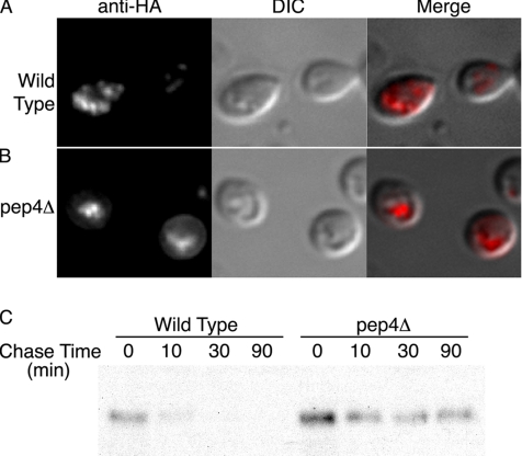
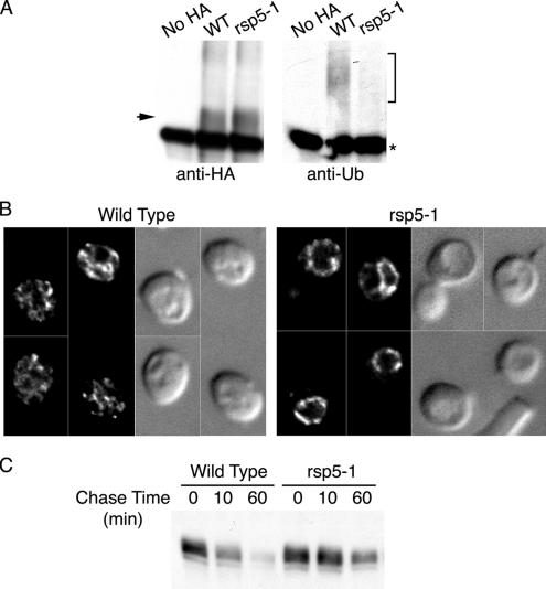
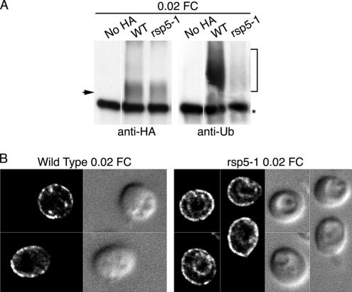
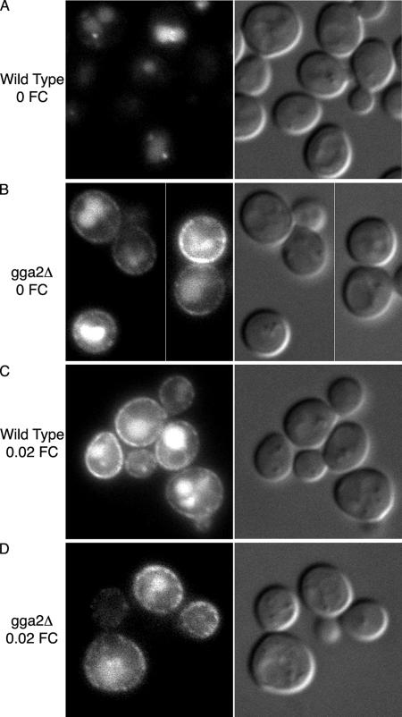
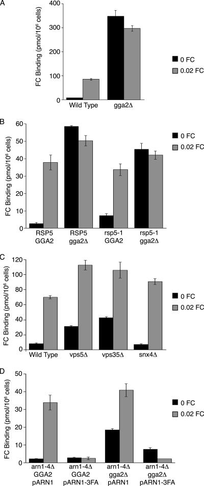
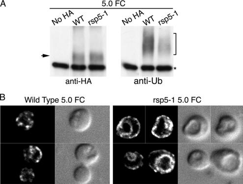
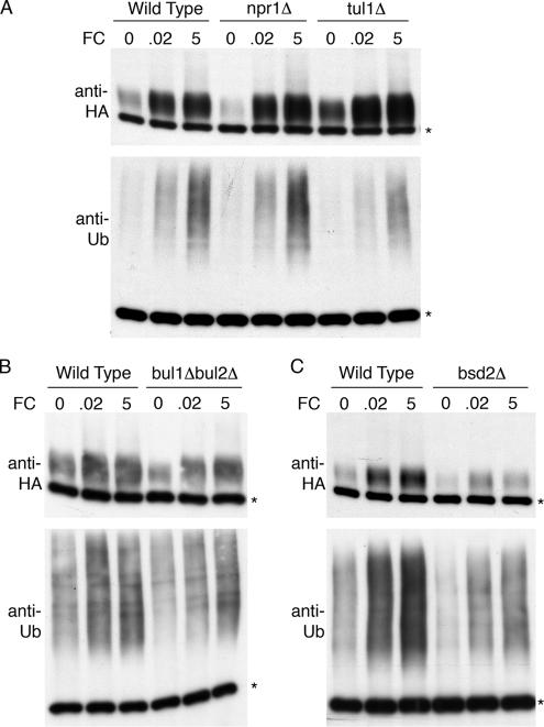
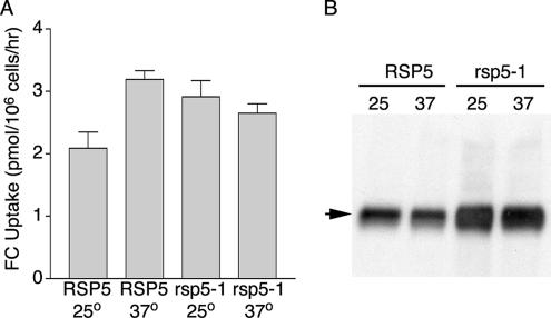
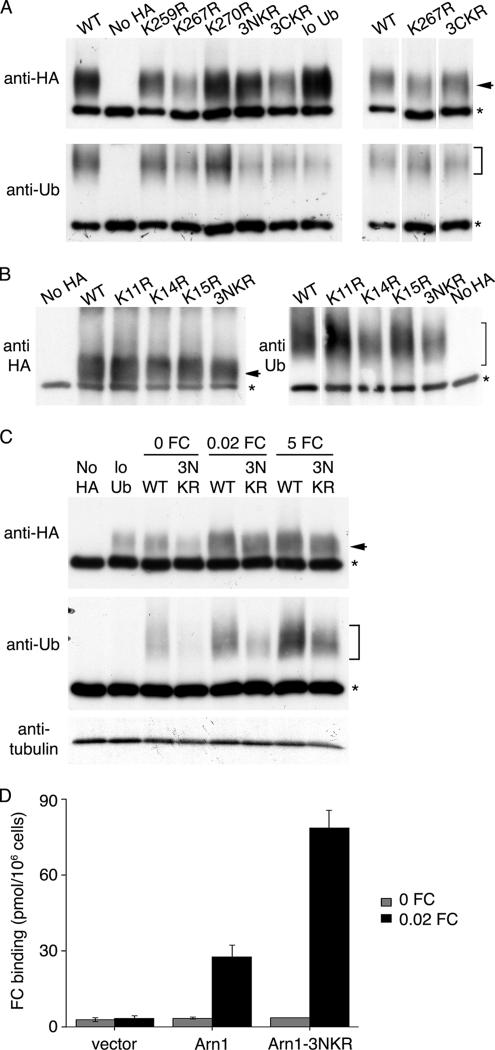
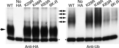
The intracellular trafficking of Arn1, a ferrichrome transporter in Saccharomyces cerevisiae, is controlled in part by the binding of ferrichrome to the transporter. In the absence of ferrichrome, Arn1 is sorted directly from the Golgi to endosomes. Ferrichrome binding triggers the redistribution of Arn1 to the plasma membrane, whereas ferrichrome transport is associated with the cycling of Arn1 between the plasma membrane and endosomes. Here, we report that the clathrin adaptor Gga2 and ubiquitination by the Rsp5 ubiquitin ligase are required for trafficking of Arn1. Gga2 was required for Golgi-to-endosomal trafficking of Arn1, which was sorted from endosomes to the vacuole for degradation. Trafficking into the vacuolar lumen was dependent on ubiquitination by Rsp5, but ubiquitination was not required for plasma membrane accumulation of Arn1 in the presence of ferrichrome. Retrograde trafficking via the retromer complex or Snx4 was also not required for plasma membrane accumulation. High concentrations of ferrichrome led to higher levels of ubiquitination of Arn1, but they did not induce degradation. Without this ubiquitination, Arn1 remained on the plasma membrane, where it was active for transport. Arn1 was preferentially modified with polyubiquitin chains on a cluster of lysine residues at the amino terminus of the transporter.


Figures

Figure 1.
Figure 1.
Vacuolar sorting and degradation of Arn1p. (A) Accumulation of Arn1p in the vacuolar lumen of pep4Δ strain. BY4742 (Wild Type) and pep4Δ strains were transformed with pArn1-HA and grown in iron-poor medium to mid-log phase before fixation and preparation for indirect immunofluorescence. Mouse monoclonal HA.11 was the primary antibody and Cy-3–conjugated donkey anti-mouse was the secondary antibody. Images are in sets of three: fluorescence on the left, differential interference contrast (DIC) in the center, and the merged image on the right. (B) Increased stability of Arn1 in pep4Δ strain. BY4742 (Wild Type) and pep4Δ strains were transformed with pMetArn1-HA and grown in SC medium without methionine to mid-log phase. Cycloheximide and methionine were added, and aliquots of cells removed at the indicated times. Cells were lysed and equivalent amounts of cellular proteins were analyzed by SDS-PAGE and immunoblotting with anti-HA antibody.
Figure 2.
Figure 2.
Requirement of Rsp5-dependent ubiquitination for vacuolar degradation of Arn1p. (A) Loss of Arn1p ubiquitination in the rsp5-1 strain. Strains YKY102 (Wild Type) and YKY103 (rsp5-1) were transformed with either pMetArn1-HA or the empty vector pRS316 (no HA) and YEp96. YEp96 expresses untagged ubiquitin from the CUP1 promoter. Transformed strains were grown at 25°C in SC medium without methionine and with 100 μM copper sulfate to induce expression of Arn1-HA and ubiquitin, respectively. Cultures were shifted to 37°C for 2 h, and then an equivalent number of cells was lysed, and the membranes were purified, detergent solubilized, and subjected to immunoprecipitation with anti-HA antibody. Immunoprecipitates were then subjected to Western blotting using an anti-HA antibody to detect Arn1-HA (left) or an anti-ubiquitin antibody to detect ubiquitinated Arn1-HA (right). The arrow indicates unmodified Arn1-HA and the bracket indicates ubiquitinated forms of Arn1. The asterisk indicates immunoglobulin heavy chain. (B) Accumulation of Arn1p at vacuolar membrane in the rsp5-1 strain. Strains YKY102 (Wild Type) and YKY103 (rsp5-1) were transformed with pArn1-HA and grown at 25°C in iron-poor medium to mid-log phase. Cultures were shifted to 37°C for 2 h before fixation and preparation for indirect immunofluorescence microscopy. Images were collected as a vertical series and analyzed by digital deconvolution. Images are in pairs with deconvolved fluorescence on the left and DIC on the right. (C) Increased stability of Arn1p in rsp5-1 strain. Strains YKY102 (Wild Type) and YKY103 (rsp5-1) were transformed with pMetArn1-HA and grown in methionine-free medium to mid-log phase at 25°C. Cultures were shifted to 37°C for 2 h before addition of cycloheximide and methionine, and aliquots of cells were removed at the indicated times. Cells were lysed and membranes were isolated and solubilized. Equivalent amounts of membrane protein were subjected to SDS-PAGE and immunoblotting with anti-HA antibody.
Figure 3.
Figure 3.
Plasma membrane trafficking of Arn1p independent of ubiquitination. (A) Loss of ubiquitination in the rsp5-1 strain. Strains YKY102 (Wild Type) and YKY103 (rsp5-1) were transformed and grown at 25°C as in Figure 2A. Cultures were shifted to 37°C with the addition of 20 nM FC for 2 h, and cells were harvested and subjected to immunoprecipitation and SDS-PAGE as in Figure 2A. (B) Ubiquitin-independent relocalization of Arn1p to the plasma membrane. Strains YKY102 (Wild Type) and YKY103 (rsp5-1) were transformed and grown as in Figure 2B except that 20 nM FC was added for 2 h before preparation for fluorescence microscopy imaging.
Figure 4.
Figure 4.
Mislocalization of Arn1-GFP in a gga2Δ strain. Wild type (A and C) and congenic gga2Δ (B and D) strains containing GFP coding sequences integrated at the carboxy terminus of ARN1 were grown overnight in iron-poor medium with either no FC (A and B) or 0.02 μM FC (C and D) added during the final two hours of growth. Epifluorescent images are on the left, DIC images on the right.
Figure 5.
Figure 5.
Surface localization of Arn1p in wild-type, gga2Δ, rsp5-1, rsp5-1 gga2Δ, and retrograde trafficking-deficient strains. (A) Mislocalization of Arn1p to cell surface in the gga2Δ strain. Strain BY4742 and the congenic gga2Δ strain were grown in iron-poor medium overnight to induce expression of Arn proteins, cultures were divided, and 0.02 μM FC was added to one aliquot for the final 2 h of growth. Cells were treated with 20 mM NaN3 and KF, harvested, washed, and mixed with [55Fe-FC] at 100 nM at 0°C for 15 min. Cells were again washed, and retained [55Fe-FC] was measured. (B) Mislocalization of Arn1p to cell surface in rsp5-1 gga2Δ strain. Congenic strains of the indicated genotypes were grown in iron medium overnight at 25°C, and then they were divided and shifted to 37°C for two additional hours of growth. FC at 0.02 μM was added to one aliquot for the final 2 h. Cells were then treated as described in A. (C) Surface localization of Arn1p in the absence of retrograde trafficking. Congenic strains of the indicated genotype were treated as described in A. (D) Failure of the Arn1p-3FA mutant to traffic to the cell surface in the gga2Δ strain. Congenic arn1–4Δ and arn1–4Δgga2Δ strains were transformed with pArn1-HA or pArn1–3FA-HA and treated as described in A. All samples were prepared in triplicate, and binding assays were repeated three times with similar results. Data from a single representative experiment are shown, error bars represent average deviation.
Figure 6.
Figure 6.
Requirement of Rsp5-dependent ubiquitination for endocytosis of Arn1p. (A) Loss of ubiquitination in the rsp5-1 strain. Strains YKY102 (Wild Type) and YKY103 (rsp5-1) were transformed and grown at 25°C as in Figure 2A. Cultures were shifted to 37°C with the addition of 5 μM FC for 2 h, and cells were harvested and subjected to immunoprecipitation and SDS-PAGE as in Figure 2A. (B) Loss of FC-induced endocytosis in the rsp5-1 strain. Strains YKY102 (Wild Type) and YKY103 (rsp5-1) were transformed and grown as in Figure 2B except that 5 μM FC was added for 2 h before preparation for fluorescence microscopy imaging.
Figure 7.
Figure 7.
Ubiquitination independent of Npr1p, Tul1p, and Rsp5p accessory proteins. Congenic strains BY4742 (Wild Type), npr1Δ, tul1Δ, and bsd2Δ (A and C), and congenic strains YPH499 (Wild Type) and FAJ75 (bul1Δbul2Δ) (B) were transformed with pMetArn1-HA and Yep96. Transformed strains were grown to mid-log phase in SC without methionine and with 100 μM copper. FC (micromolar) was added at the indicated concentrations for the last 2 h. An equivalent number of cells were lysed, the membranes were purified and subjected to immunoprecipitation with anti-HA antibody. Immunoprecipitates were then subjected to Western blotting using anti-HA antibodies to detect Arn1-HA (top) or anti-ubiquitin antibodies (P4D1) to detect ubiquitinated Arn1 (bottom).
Figure 8.
Figure 8.
Transport of ferrichrome in absence of ubiquitination. Congenic strains YKY104 (RSP5+ fet3Δ) and YKY105 (rsp5-1 fet3Δ) were transformed with pArn1-HA(ADE) and were grown at 25°C in iron-poor medium. Cultures were divided and shifted to 37°C for 1 h and divided again for the measurement of uptake of [55Fe] FC (A) and for Western blotting (B). (A) Uptake of FC in the rsp5-1 strain. Uptake assays were performed using four samples for each data point and were repeated three times; data from a representative experiment are shown. (B) Increased levels of Arn1p-HA in the rsp5-1 strain. Cells were lysed and membranes were isolated and solubilized. Equivalent amounts of membrane protein were subjected to SDS-PAGE and immunoblotting with anti-HA antibody. Arrow indicates Arn1p-HA.
Figure 9.
Figure 9.
Identification of amino-terminal lysine residues as targets of ubiquitination. (A) Preferential ubiquitination of amino-terminal lysines. (B) Identification of lysine 14 of Arn1p as primary target of ubiquitination. (C) Targeting of amino-terminal lysines at all ferrichrome concentrations. YPH499 was transformed with YEp96 and a plasmid containing either wild-type Arn1-HA (pMetArn1-HA, WT) or mutant forms of Arn1-HA (K11R, K14R, K15R, K259R, K267R, K270R, 3NKR, or 3CKR). YPH499 transformed with pRS316 and YEp96 was used as the no-HA control. YPH499 integrated with pRS404 and transformed with pMetArn1-HA was used as the low-Ub control. Transformants were grown in SC without methionine and with 100 μM copper. FC was added at the last hour to the final concentration of 5 μM (A and B) or as indicated (C). Equivalent numbers of cells were harvested from each culture. Cells were lysed and membranes were purified, detergent solubilized, and subjected to immunoprecipitation with anti-HA antibody. Immunoprecipitates were then subjected to Western blotting using anti-HA antibodies to detect Arn1-HA (top) or anti-Ub antibodies to detect ubiquitinated Arn1 (bottom). Equivalent aliquots of the solubilized membranes used for immunoprecipitations in C were subjected to Western blotting with anti-tubulin antibody as a control. (D) Absence of surface mislocalization in Arn1-3NKR. The strain CWY101, from which all ARN transporters have been deleted, was transformed with pMetArn1-HA, pMetArn1-3NKR-HA, or the empty vector pRS316. Transformants were grown in SC medium, cultures were divided, and 0.02 μM FC was added to one aliquot for the final 2 h of growth. Cells were treated with 20 mM NaN3 and KF, harvested, washed, and mixed with [55Fe-FC] at 100 nM at 0°C for 15 min. Cells were again washed and retained [55Fe-FC] was measured. All samples were prepared in triplicate, and binding assays were repeated three times with similar results. Data from a single representative experiment are shown; error bars represent average deviation.
Figure 10.
Figure 10.
Polyubiquitination of Arn1p at multiple sites in the presence of ferrichrome. YPH499 was transformed with pMetArn1-HA and a plasmid containing either wild-type ubiquitin (YEp96, WT) or mutant forms of ubiquitin (K29R, K48R, K63R, 6K-R). YPH499 transformed with pRS316 and Yep96 was used as the no-HA control. Transformed strains were grown in synthetic complete medium without methionine and with 100 μM copper. Ferrichrome at 5 μM was added in the last hour. Equivalent numbers of cells were harvested from each culture and treated as described in Figure 7 legend. Single arrow indicates unmodified Arn1p-HA. Double-headed arrows indicate bands corresponding to ubiquitinated species of Arn1p-HA.

Similar articles

Cited by

References

    1. Abe F., Horikoshi K. Tryptophan permease gene TAT2 confers high-pressure growth in Saccharomyces cerevisiae. Mol. Cell Biol. 2000;20:8093–8102. - PMC - PubMed
    1. Abe F., Iida H. Pressure-induced differential regulation of the two tryptophan permeases Tat1 and Tat2 by ubiquitin ligase Rsp5 and its binding proteins, Bul1 and Bul2. Mol. Cell Biol. 2003;23:7566–7584. - PMC - PubMed
    1. Babst M., Sato T. K., Banta L. M., Emr S. D. Endosomal transport function in yeast requires a novel AAA-type ATPase, Vps4p. EMBO J. 1997;16:1820–1831. - PMC - PubMed
    1. Barriere H., Nemes C., Lechardeur D., Khan-Mohammad M., Fruh K., Lukacs G. L. Molecular basis of oligoubiquitin-dependent internalization of membrane proteins in mammalian cells. Traffic. 2006;7:282–297. - PubMed
    1. Bilodeau P. S., Winistorfer S. C., Allaman M. M., Surendhran K., Kearney W. R., Robertson A. D., Piper R. C. The GAT domains of clathrin-associated GGA proteins have two ubiquitin binding motifs. J. Biol. Chem. 2004;279:54808–54816. - PMC - PubMed

Publication types

MeSH terms