. 2007 Mar 8:7:14.
doi: 10.1186/1472-6750-7-14.

Single chain Fab (scFab) fragment

Affiliations

Single chain Fab (scFab) fragment

Michael Hust et al. BMC Biotechnol. .

Abstract

Background: The connection of the variable part of the heavy chain (VH) and and the variable part of the light chain (VL) by a peptide linker to form a consecutive polypeptide chain (single chain antibody, scFv) was a breakthrough for the functional production of antibody fragments in Escherichia coli. Being double the size of fragment variable (Fv) fragments and requiring assembly of two independent polypeptide chains, functional Fab fragments are usually produced with significantly lower yields in E. coli. An antibody design combining stability and assay compatibility of the fragment antigen binding (Fab) with high level bacterial expression of single chain Fv fragments would be desirable. The desired antibody fragment should be both suitable for expression as soluble antibody in E. coli and antibody phage display.

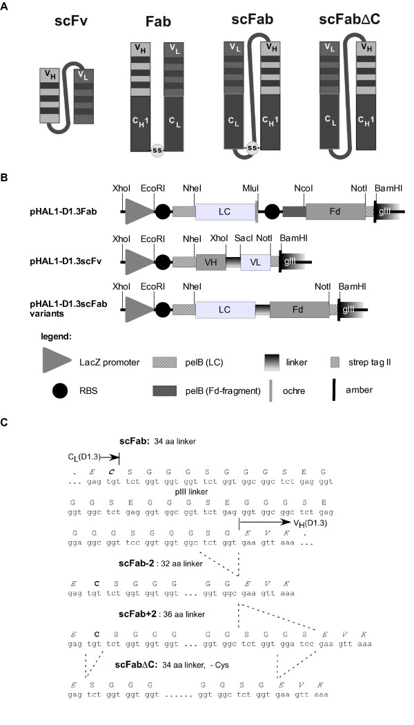
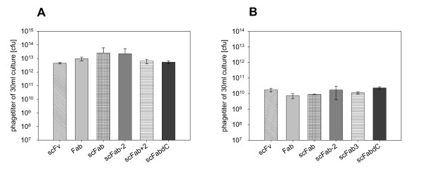
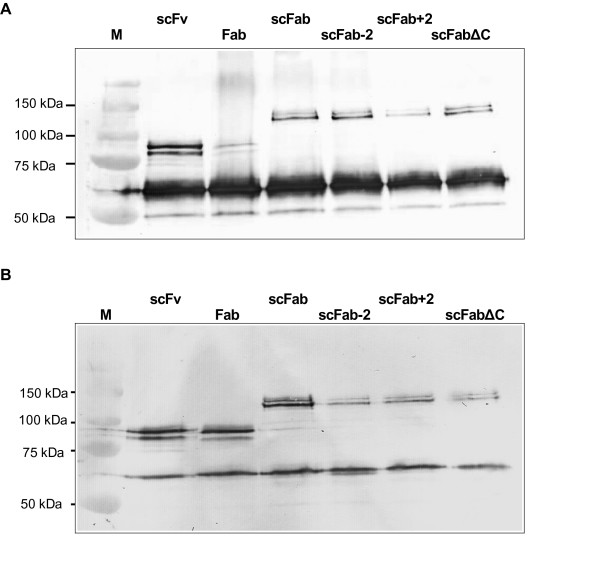
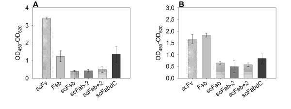
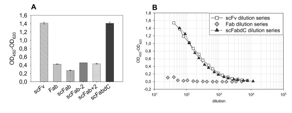
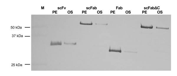
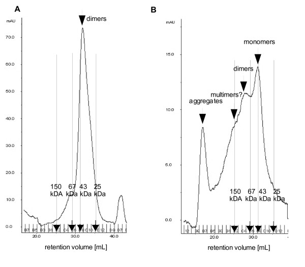
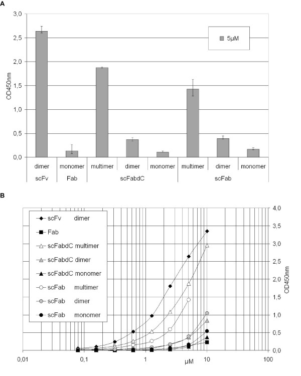
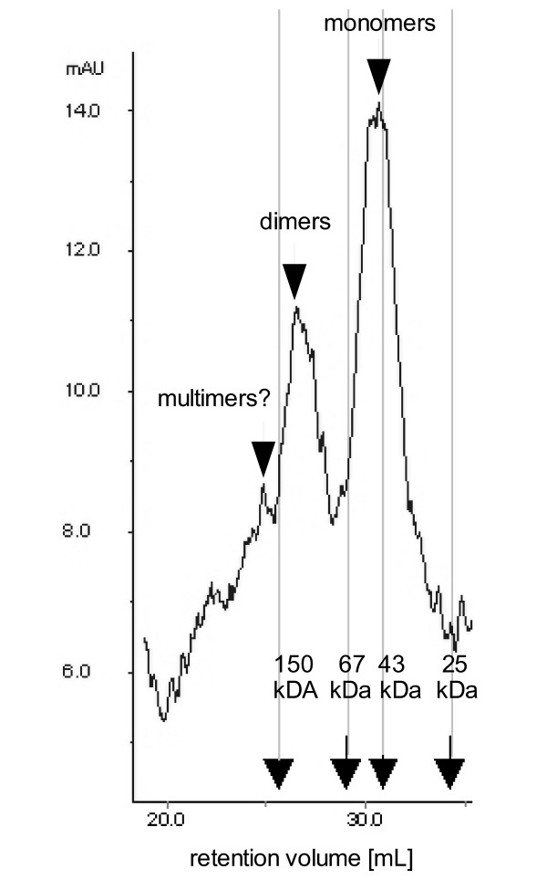
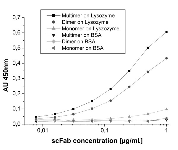
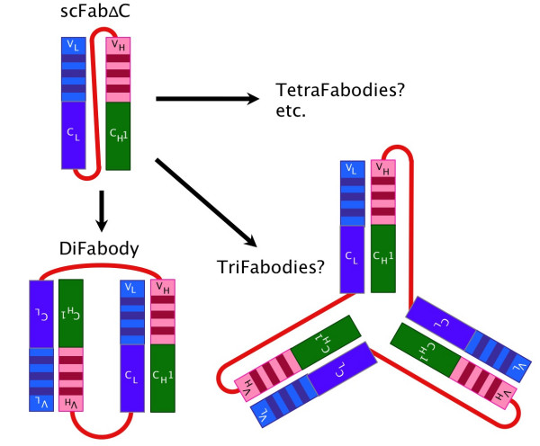
Results: Here, we demonstrate that the introduction of a polypeptide linker between the fragment difficult (Fd) and the light chain (LC), resulting in the formation of a single chain Fab fragment (scFab), can lead to improved production of functional molecules. We tested the impact of various linker designs and modifications of the constant regions on both phage display efficiency and the yield of soluble antibody fragments. A scFab variant without cysteins (scFabDeltaC) connecting the constant part 1 of the heavy chain (CH1) and the constant part of the light chain (CL) were best suited for phage display and production of soluble antibody fragments. Beside the expression system E. coli, the new antibody format was also expressed in Pichia pastoris. Monovalent and divalent fragments (DiFabodies) as well as multimers were characterised.

Conclusion: A new antibody design offers the generation of bivalent Fab derivates for antibody phage display and production of soluble antibody fragments. This antibody format is of particular value for high throughput proteome binder generation projects, due to the avidity effect and the possible use of common standard sera for detection.


Figures

Figure 1
Figure 1
Vectors constructed for this study. A illustration of the different antibody formats, B the pHAL1-D1.3 vectors, C linker sequences of the scFab variants. Abbreviations: lacZ promoter: promoter of the bacterial lac operon; RBS: ribosome binding site; pelB: signal peptide sequence of bacterial pectate lyase, mediating secretion into the periplasmic space; VH: variable fragment of the heavy chain; LC: light chain; ochre: ochre stop codon; amber: amber stop codon; strep-tag II: synthetic tag binding to streptactin. The elements of the inserts are not drawn to scale.
Figure 2
Figure 2
Comparison of antibody phage titres (cfu) obtained from 30 mL of culture of six different D1.3 antibody fragment variants. Packaged A with M13K07, B with Hyperphage. Mean value and standard deviations are from three completely independent experiments.
Figure 3
Figure 3
Immunoblot of antibody phage presenting different formats, produced with either M13K07 or Hyperphage. A 1 × 10¹¹ D1.3 antibody phage produced with M13K07 were separated, B 3 × 10⁸ D1.3 antibody phage produced with Hyperphage were separated. Phage were separated on a reducing 10 % SDS-PAGE, pIII was detected using mouse mAb anti-pIII.
Figure 4
Figure 4
Antigen binding phage ELISA of phage presenting D1.3 antibody fragment variants. A 5 × 10⁸ phage produced with M13K07, B 107 phage produced with Hyperphage. Mean values and standard deviations of three completely independent experiments are given. The absorbance of scattered light at 620 nm was subtracted from the absorbance at 450 nm. The background signal of the antigen incubated with the detection antibody mAb anti-M13 HRP was subtracted. Antigen: 100 ng/well lysozyme. The D1.3 antibody phage were detected using mAb anti-M13 conjugated with HRP.
Figure 5
Figure 5
Antigen binding ELISA of soluble antibody fragments produced in E. coli using pHAL1 with 1 μg/well lysozyme coated per well. A 20 μL periplasmic fractions from production in MTPs of scFv, Fab and four scFab constructs were applied, the D1.3 antibodies were detected using mouse mAb anti-Strep-Tag (1:10000), B serial dilutions of Protein L purified equimolar amounts of scFv, Fab and scFabΔC were applied, D1.3 antibody fragments were detected using Protein L conjugated to HRP (1:10000)
Figure 6
Figure 6
Immunoblot of soluble antibody fragments produced using the E. coli expression vector pOPE101. 20 μL aliquots of E. coli periplasmic fractions were separated on reducing 10% SDS-PAGE, pIII was detected using mouse mAb anti-myc tag and goat anti-mouse IgG AP. Abbreviations: PE: periplasmic fraction; OS: osmotic shock preparation.
Figure 7
Figure 7
Size exclusion chromatography analysis of soluble antibody fragments produced using the E. coli expression vector pOPE101. 50 μg purified antibody fragments of the A scFv, or B scFabΔC format were separated on superdex 200. Calibration was done with Chymotrypsinogen (25 kDa), Ovalbumin (43 kDa), Albumin (67 kDa) and IgG (150 kDa).
Figure 8
Figure 8
Antigen binding ELISA of soluble antibody fragments produced in E. coli using pOPE101 with 1 μg/well lysozyme coated per well. A 5 μM gelfiltration purified fractions of scFv, Fab and four scFab constructs were applied per well, D1.3 antibody fragments were detected using mAb anti-myc tag (1:25), the ELISA measurements were done in triplicate. B ELISA signals of serial dilutions of the different fractions of the antibody fragments.
Figure 9
Figure 9
Size exclusion analysis of soluble antibody fragments produced in P. pastoris. 500 μg purified antibody fragments of the scFabΔC format were separated on Superdex 200.
Figure 10
Figure 10
Antigen binding ELISA of soluble antibody fragments produced in P. pastoris with 100 ng/well lysozyme or BSA coated per well. A serial dilution of gel filtration purified fractions of scFabΔC ranging from 8 ng/mL to 1 μg/mL were applied, the D1.3 antibody fragments were detected using Protein L HRP conjugate (1:10000). Measurements were done in duplicate.
Figure 11
Figure 11
Illustration of scFabΔC and the multimerisation forms DiFabody and TriFabody.

Similar articles

Cited by

References

    1. Skerra A, Plückthun A. Assembly of a functional immunoglobulin Fv fragment in Escherichia coli. Science. 1988;240:1038–1041. doi: 10.1126/science.3285470. - DOI - PubMed
    1. Bird RE, Hardman KD, Jacobsen JW, Johnson S, Kaufman BM, Lee SM, Lee T, Pope SH, Riordan GS, Whitlow M. Single-chain antigen-binding proteins. Science. 1988;242:423–426. doi: 10.1126/science.3140379. - DOI - PubMed
    1. Huston JS, Levinson D, Mudgett HM, Tai MS, Novotny J, Margolies MN, Ridge RJ, Bruccoloreri RE, Haber E, Crea R, Oppermann H. Protein engineering of antibody binding sites: recovery of specific activity in an anti-digosin single-chain Fv analogue produced in Escherichia coli. Proc Natl Acad Sci USA. 1988;85:5879–5883. doi: 10.1073/pnas.85.16.5879. - DOI - PMC - PubMed
    1. Marks JD, Hoogenboom HR, Griffiths AD, Winter G. Molecular evolution of proteins on filamentous phage. J Biol Chem. 1992;267:16007–16010. - PubMed
    1. Bird RE, Walker BW. Single chain variable regions. Trends Biotech. 1991;9:132–137. doi: 10.1016/0167-7799(91)90044-I. - DOI - PubMed

Publication types

MeSH terms

LinkOut - more resources