. 2008 Jul;29(7):1002-10.
doi: 10.1016/j.neurobiolaging.2007.02.009. Epub 2007 Mar 7.

Brain and behavior changes in 12-month-old Tg2576 and nontransgenic mice exposed to anesthetics

Affiliations

Brain and behavior changes in 12-month-old Tg2576 and nontransgenic mice exposed to anesthetics

Shannon L Bianchi et al. Neurobiol Aging. 2008 Jul.

Abstract

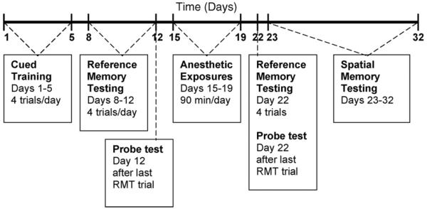
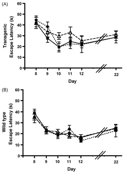
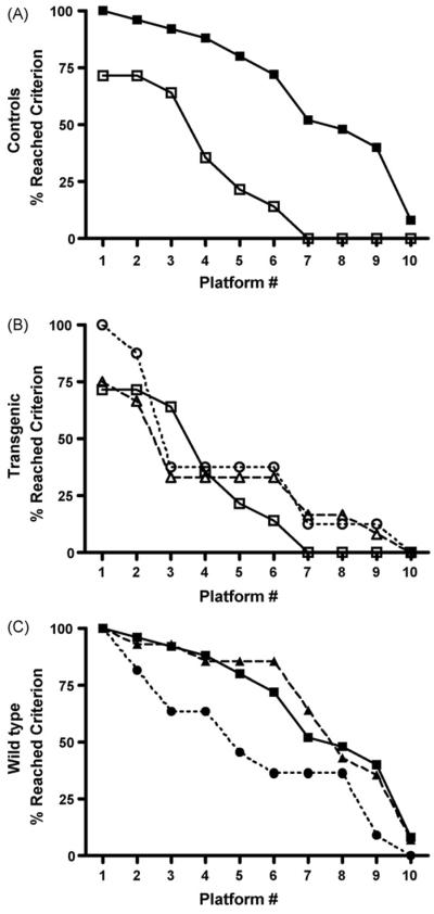
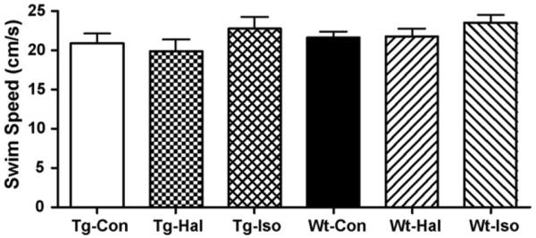
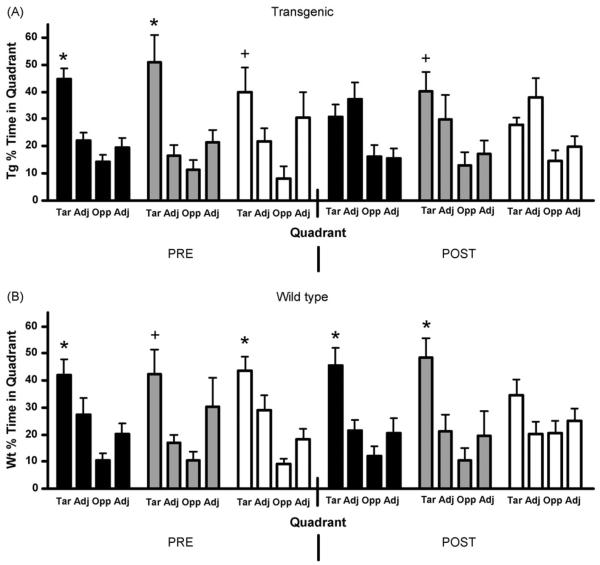
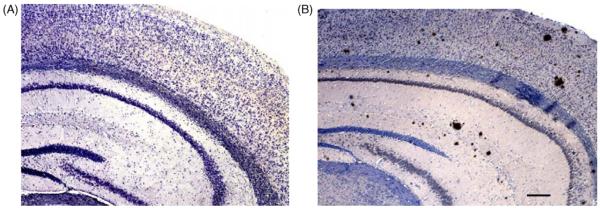
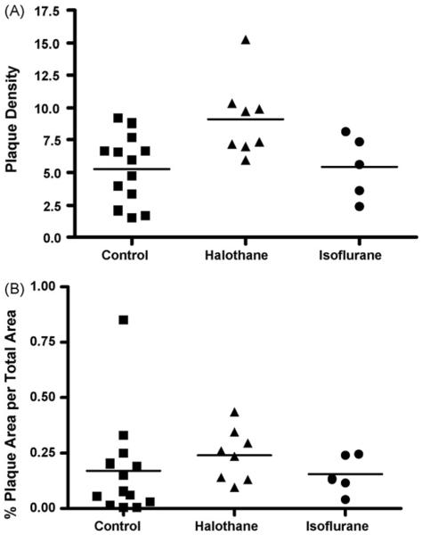
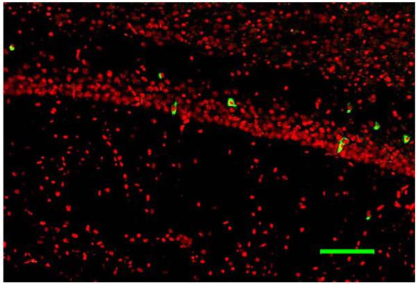
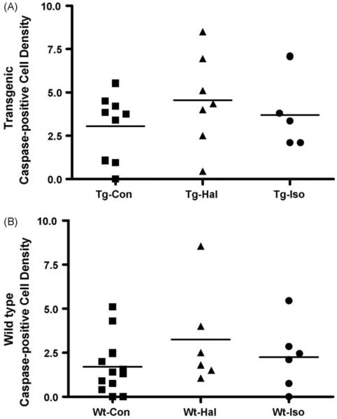
Inhaled anesthetics have been shown to increase the aggregation of amyloid beta in vitro through the stabilization of intermediate toxic oligomers, which are thought to contribute to neurocognitive dysfunction in Alzheimer's disease. Inhaled anesthetics may escalate cognitive dysfunction through enhancement of these intermediate oligomer concentrations. We intermittently exposed 12-month-old Tg2576 transgenic mice and nontransgenic littermates to isoflurane and halothane for 5 days. Cognitive function was measured before and after anesthetic exposures using the Morris Water Maze; amyloid beta plaque burden and caspase-3 mediated apoptosis were quantified by immunohistochemistry. At 12 months of age, anesthetic exposure did not further enhance cognitive decline in the transgenic mice. Immunohistochemistry, however, revealed that the halothane-exposed Tg2576 mice had more amyloidopathy than the isoflurane treated mice or the nonexposed transgenic mice. Isoflurane exposure impaired cognitive function in the nontransgenic mice, implying an alternative pathway for neurodegeneration. These findings indicate that inhaled anesthetics influence cognition and amyloidogenesis, but that the mechanistic relationship remains unclear.


Figures

Fig. 1
Fig. 1
Schematic time-line of the experimental paradigm. RMT= reference memory testing.
Fig. 2
Fig. 2
Escape latencies from the reference memory testing for transgenic (A) and wild-type mice (B). No changes were found in the escape latencies of any group before and after the exposures. The x-axis, days, corresponds to the testing paradigm in Fig. 1 for pre-exposures (days 8–12) and post-exposure (day 22). Black squares and solid line, controls; open triangles and dashed line, halothane-exposed; black circles and small dashed lines, isoflurane-exposed (Tg-Hal, n = 12; Tg-Iso, n = 5; Tg-Con, n = 12; Wt-Hal, n = 14; Wt-Iso, n = 7; Wt-Con, n = 24).
Fig. 3
Fig. 3
The percent of transgenic and wild-type mice that reached criterion in the spatial memory testing after anesthetic exposures. The x-axis represents the number of different hidden platform locations reached by the mice during the 10 days of testing. (A) Control mice: wild-type (solid squares, solid line) (n = 25) compared to transgenic (open squares, solid line) (n = 14) (p < 0.0001). (B) Transgenic mice: controls (open squares, solid line) (n = 14), isoflurane exposed (open circles, dashed line) (n=5) and halothane-exposed (open triangles, dotted line) (n = 12) (p > 0.05). No significant difference was detected between any transgenic group. (C) Wild-type mice: controls (solid squares, solid line) (n = 25), isoflurane exposed (solid circles, dashed line) (n = 11) and halothane-exposed (solid triangles, dotted line) (n = 14). Performance in the isoflurane group was significantly different from control, but not in the halothane group.
Fig. 4
Fig. 4
The average swim speeds of transgenic (Tg) and wild-type (Wt) mice during the spatial memory testing after exposures to halothane (Hal) or isoflurane (Iso). No significant differences in swim speed were observed (p = 0.4037) (Tg-Con, n = 14; Tg-Hal, n = 12; Tg-Iso, n = 5; Wt-Con, n = 25; Wt-Hal, n = 14; Wt-Iso, n = 11).
Fig. 5
Fig. 5
The percent of total time that mice spent in each quadrant during pre- and post-exposure probe tests for controls (black), halothane (grey) and isoflurane (white) exposed mice. (A) Transgenic mice, prior to anesthetic exposure, spent more time in the target quadrant than the other quadrants. However, after the exposures, only the halothane group spent significantly more time in the target quadrant. (B) Wild-type mice significantly preferred the target over adjacent or opposite quadrants both before and after anesthetic exposures, except for the post-exposure isoflurane group (*, p < 0.05 target vs. opposite and adjacent quadrants; +, p < 0.05 target vs. opposite quadrant only) (Tar = target, Adj = adjacent, Opp = opposite) (Tg-Con, n = 10; Tg-Hal, n = 6; Tg-Iso, n = 5; Wt-Con, n = 10; Wt-Hal, n = 6; Wt-Iso, n = 7).
Fig. 6
Fig. 6
Micrographs of a wild-type mouse brain (A) and transgenic brain (B). Variously sized amyloid plaques were found in the cortex, CA1 region and dentate gyrus in the transgenic brain at 12 months of age (B). Scale bar = 180 μm.
Fig. 7
Fig. 7
The plaque load in the Tg2576 mouse cortex, CA1 and dentate gyrus combined. (A) The density of plaques in controls and anesthetic exposedmice. The halothane-exposed Tg2576 mice had a significantly increased plaque burden compared to control Tg2576 mice, p < 0.05. (B) The percent area occupied by plaque normalized to area. No significant differences were detected in the anesthetic exposed groups (Con, n = 13; Hal, n = 8; Iso, n = 5).
Fig. 8
Fig. 8
Caspase-3 immunohistochemistry of the CA1 region of the hippocampus in a transgenic mouse exposed to isoflurane. Caspase-3 positive cells are green. Scale bar = 50μm.
Fig. 9
Fig. 9
The density of caspase-3 positive cells in the cortex, CA1 and dentate gyrus combined. Overall, the density of caspase-positive cells is significantly greater in all transgenic mice than wild-type mice (p < 0.02), but an anesthetic effect could not be demonstrated (Wt-Con, n = 12; Wt-Hal n = 6; Wt- Iso n = 6; Tg-Con, n = 9; Tg-Hal, n = 7; Tg-Iso, n = 5).

References

    1. Abildstrom H, Rasmussen LS, Rentowl P, Hanning CD, Rasmussen H, Kristensen PA, Moller JT. Cognitive dysfunction 1–2 years after non-cardiac surgery in the elderly. ISPOCD group. International Study of Post-Operative Cognitive Dysfunction. Acta Anaesthesiol. Scand. 2000;44(10):1246–1251. - PubMed
    1. Ancelin ML, de Roquefeuil G, Ledesert B, Bonnel F, Cheminal JC, Ritchie K. Exposure to anaesthetic agents, cognitive functioning and depressive symptomatology in the elderly. Br. J. Psychiatry. 2001;178:360–366. - PubMed
    1. Avila J. Tau phosphorylation and aggregation in Alzheimer's disease pathology. FEBS Lett. 2006;580(12):2922–2927. - PubMed
    1. Bedford PD. Adverse cerebral effects of anesthesia on old people. Lancet. 1955;ii:259–263. - PubMed
    1. Billings LM, Oddo S, Green KN, McGaugh JL, LaFerla FM. Intraneuronal Abeta causes the onset of early Alzheimer's disease-related cognitive deficits in transgenic mice. Neuron. 2005;45(5):675–688. - PubMed

Publication types

MeSH terms