

Altered orbitofrontal sulcogyral pattern in schizophrenia
- PMID: 17347256
- PMCID: PMC2768130
- DOI: 10.1093/brain/awm007
Altered orbitofrontal sulcogyral pattern in schizophrenia
Abstract
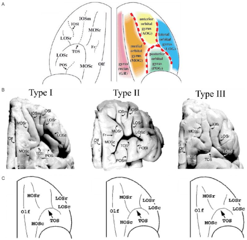
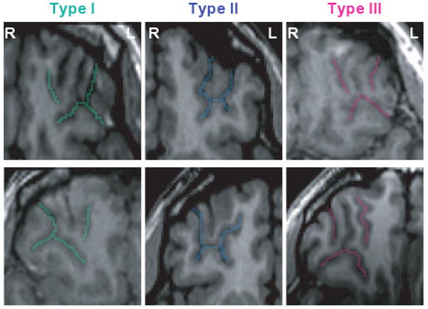
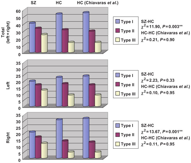
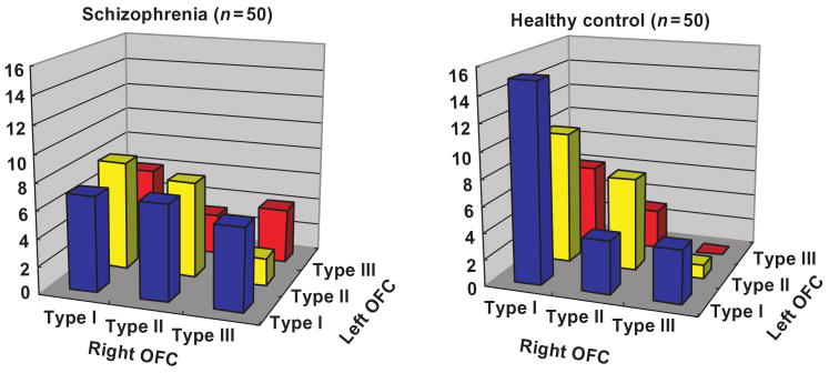
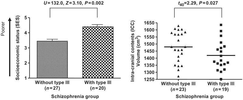
Orbitofrontal alteration in schizophrenia has not been well characterized, likely due to marked anatomical variability. To investigate the presence of such alterations, we evaluated the sulcogyral pattern of this 'H-shaped' sulcus. Fifty patients with schizophrenia (100 hemispheres) and 50 age- and gender-matched control subjects (100 hemispheres) were evaluated using 3D high-spatial resolution MRI. Based on a previous study by Chiavaras and Petrides (2000), the sulcogyral pattern of the 'H-shaped' sulcus, which forms the boundaries of major orbitofrontal gyri, was classified into three types (Type I, II and III, in order of frequency) within each hemisphere. Chi-square analysis was performed to compare the sulcogyral pattern, and categorical regression was applied to investigate clinical/cognitive associations. The control data replicated the orbitofrontal sulcogyral pattern reported by Chiavaras and Petrides (P = 0.90-0.95), where the distribution was significantly different between the left and right hemisphere (Type I: right > left, Type II, III: left > right, chi2 = 6.41, P = 0.041). For schizophrenics, the distribution differed significantly from controls (chi2 = 11.90, P = 0.003), especially in the right hemisphere (chi2 = 13.67, P = 0.001). Moreover, the asymmetry observed in controls was not present in schizophrenia (chi2 = 0.13, P = 0.94). Specifically, the most frequent Type I expression was decreased and the rarest Type III expression was increased in schizophrenia, relative to controls. Furthermore, patients with Type III expression in any hemisphere evinced poorer socioeconomic status, poorer cognitive function, more severe symptoms and impulsivity, compared to patients without Type III expression. In contrast, patients with Type I in any hemisphere showed better cognitive function and milder symptoms compared to patients without Type I. Structurally, patients with Type III had significantly smaller intra-cranial contents (ICC) volumes than did patients without Type III (t(40) = 2.29, P = 0.027). The present study provides evidence of altered distribution of orbitofrontal sulcogyral pattern in schizophrenia, possibly reflecting a neurodevelopmental aberration in schizophrenia. Such altered sulcogyral pattern is unlikely to be due to secondary effects of the illness such as medication. Moreover, the structural association between Type III and small ICC volume, observed in the patient group, may suggest that Type III expression could be part of a systematic neurodevelopmental alteration, given that the small ICC volume could reflect early reduction of cranial growth driven by brain growth. The observed contrasting association of Type III expression with poorer outcome, and that of Type I expression with better outcome, further suggests clinical heterogeneity, and possible differences in treatment responsiveness in schizophrenia.
Figures
References
-
- Armstrong E, Schleicher A, Omran H, Curtis M, Zilles K. The ontogeny of human gyrification. Cereb Cortex. 1995;5:56–63. - PubMed
-
- Baare WF, Hulshoff Pol HE, Hijman R, Mali WP, Viergever MA, Kahn RS. Volumetric analysis of frontal lobe regions in schizophrenia: relation to cognitive function and symptomatology. Biol Psychiatry. 1999;45:1597–605. - PubMed
-
- Bartley AJ, Jones DW, Weinberger DR. Genetic variability of human brain size and cortical gyral patterns. Brain. 1997;120:257–69. - PubMed
-
- Bleuler E. In: Dementia praecox of the group of schizophrenias. Zinkin J, translator. New York, NY: International Universities Press; 1950. 1911.
-
- Bouix S, Ungar L, Dickey CC, McCarley RW, Shenton ME. Evaluating automatic brain tissue classifiers. International Conference on Medical Image Computing and Computer-Assisted Intervention; St. Malo, France. 2004. pp. 1038–9.
Publication types
MeSH terms
Grants and funding
LinkOut - more resources
Full Text Sources
Medical
