. 2007 Jun 1;581(Pt 2):829-39.
doi: 10.1113/jphysiol.2007.129049. Epub 2007 Mar 8.

Additive protective effects of the addition of lactic acid and adrenaline on excitability and force in isolated rat skeletal muscle depressed by elevated extracellular K+

Affiliations

Additive protective effects of the addition of lactic acid and adrenaline on excitability and force in isolated rat skeletal muscle depressed by elevated extracellular K+

Frank Vincenzo de Paoli et al. J Physiol. .

Abstract

During strenuous exercise, extracellular K(+) ([K(+)](o)) is increased, which potentially can reduce muscle excitability and force production. In addition, exercise leads to accumulation of lactate and H(+) and increased levels of circulating catecholamines. Individually, reduced pH and increased catecholamines have been shown to counteract the depressing effect of elevated K(+). This study examines (i) whether the effects of addition of lactic acid and adrenaline on the excitability of isolated muscles are caused by separate mechanisms and are additive and (ii) whether the effect of adding lactic acid or increasing CO(2) is related to a reduction of intra- or extracellular pH. Rat soleus muscles were incubated at a [K(+)](o) of 15 mM, which reduced tetanic force by 85%. Subsequent addition of 20 mM lactic acid or 10(-5) M adrenaline led to a small recovery of force, but when added together induced an almost complete force recovery. Compound action potentials showed that the force recovery was associated with recovery of muscle excitability. The improved excitability after addition of adrenaline was associated with increased Na(+)-K(+) pump activity resulting in hyperpolarization and an increase in the chemical Na(+) gradient. In contrast, addition of lactic acid had no effect on the membrane potential or the Na(+)-K(+) pump activity, but most likely increased excitability via a reduction in intracellular pH. It is concluded that the protective effects of acidosis and adrenaline on muscle excitability and force took place via different mechanisms and were additive. The results suggest that circulating catecholamines and development of acidosis during exercise may improve the tolerance of muscles to elevated [K(+)](o).


Figures
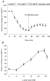
Figure 1
Figure 1. Effect of lactic acid on tetanic force in muscles at 12.5 mM K+
Tetanic contractions were elicited by applying brief trains (2 s) of pulses at 30 Hz trains were given every 10 min. After control contractions in 4 mm K+, [K+]o was increased to 12.5 mm. After a futher 70 min at 12.5 mm K+, 5 to 26 mm lactic acid was added and the incubation was continued steady-state force was obtained. A, time course of the effect of 10 mm lactic acid on force in muscles at 12.5 mm K+. B, maximum force obtained after addition of the indicated concentrations of lactic acid. Symbols show means ±s.e.m. of 6–10 muscles.
Figure 2
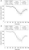
Figure 2. Effects of lactic acid and adrenaline on tetanic force in muscles incubated at 15 mM K+
Tetanic contractions were elicited every 10 min by applying brief trains (2 s) of pulses at 30 Hz. After control contractions in 4 mm K+, [K+]o was increased to 15 mm. At t = 100 min, 20 mm lactic acid (buffer pH ∼6.8; ^ or 10−5m adrenaline (buffer pH ∼7.4; •) was added. At t = 150 min adrenaline was added to the muscles already exposed to lactic acid. Symbols show means ±s.e.m. of 3–8 muscles.
Figure 3
Figure 3. Effect of lactic acid and adrenaline on the relation between [K+]o and tetanic force
Experiments were done as depicted in Fig. 2, using concentrations of extracellular K+ from 4 to 17 mm. For control muscles and muscles exposed to lactic acid, the forces shown are steady-state forces at the indicated [K+]o. For adrenaline the recovery of force was temporary (Fig. 2). For that reason the largest force production which was observed 10 min after addition adrenaline was used. □, control muscles, buffer pH ∼7.4 (n = 4–6); ▴, 20 mm lactic acid added, buffer pH ∼6.8 (n = 8–10); ^, 10−5m adrenaline added, buffer pH ∼7.4 (n = 6); ▪, 20 mm lactic and 10−5m adrenaline added, buffer pH ∼6.8 (n = 8). Continuous lines represent Bolzmann curves fitted to data. Symbols represent means ±s.e.m.
Figure 4
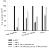
Figure 4. Effects of high [K+]o, lactic acid and adrenaline on tetanic force and M-wave parameters
M-wave recordings obtained from a muscle at 4 mm K+, after 70 min incubation at 12 mm K+, 50 min following the addition of lactic acid and 10 min after further addition of adrenaline. Values obtained during the experiment are average of 23 M-wave recordings obtained during a 1.5 s, 30 Hz tetanic train. n = 5. Bars show means ±s.e.m.
Figure 5
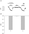
Figure 5. Effect of lactic acid and adrenaline on pHi in muscles at 11 mM K+
A, typical recordings of pHi from a muscle exposed to first lactic acid, then adrenaline and finally to both lactic acid and adrenaline. Bars indicate time of presence of the indicated compounds. In the last experiment, lactic acid was added 4 min before the addition of adrenaline. Between each treatment, muscles were washed 3 times in control buffer with 11 mm K+. B, change in pHi (mean ±s.e.m., n = 4–7) induced by addition of lactic acid, adrenaline or both lactic acid and adreline, as illustrated in A. The reduction in pHi was calculated from the difference between the lowest value for pHi obtained during 10–30 min exposure to the indicated compound and the average of the values for pHi obtained before and after the exposure.
Figure 6
Figure 6. Effects of increased HCO3 and CO2 on force in muscles depressed by high K+
Effect on tetanic force in muscles incubated at 4 or 11 mm K+ of increasing CO2 from 5 to 15% with (A) or without (B) a simultaneous increased in HCO3 from 25 to 70 mm. Tetanic contractions were elicited every 10 min by applying 2 s trains of pulses at 30 Hz. Muscles were incubated in KR buffer with the modifications indicated in the bars. Buffers with 70 mm HCO3 were made by substituting 45 mm of NaCl with 45 mm NaHCO3. Since this reduced the buffer Cl concentration by 45 mm, 45 mm NaCl was replaced with 45 mm of sodium methanesulphonate in the control buffers with only 25 mm NaHCO3 whereby the concentration of buffer Cl was kept constant throughout all the experiments.

Comment in

Similar articles

Cited by

References

    1. Albuquerque EX, Thesleff S. A comparative study of membrane properties of innervated and chronically denervated fast and slow skeletal muscles of the rat. Acta Physiol Scand. 1968;73:471–480. - PubMed
    1. Andersen SL, Clausen T. Calcitonin gene-related peptide stimulates active Na+-K+ transport in rat soleus muscle. Am J Physiol. 1993;268:C1528–C1536. - PubMed
    1. Bouclin R, Charbonneau E, Renaud JM. Na+ and K+ effect on contractility of frog sartorius muscle: implication for the mechanism of fatigue. Am J Physiol. 1995;268:C1528–C1536. - PubMed
    1. Boyd IA, Martin AR. Membrane constants of mammalian fibres. J Physiol. 1959;147:450–457. - PMC - PubMed
    1. Buchanan R, Nielsen OB, Clausen T. Excitation- and β2-agonist-induced activation of the Na+-K+ pump in rat soleus muscle. J Physiol. 2002;545:229–240. - PMC - PubMed

Publication types

MeSH terms