. 2007 Jun 1;581(Pt 2):553-65.
doi: 10.1113/jphysiol.2006.126417. Epub 2007 Mar 8.

Auto-inhibition of rat parallel fibre-Purkinje cell synapses by activity-dependent adenosine release

Affiliations

Auto-inhibition of rat parallel fibre-Purkinje cell synapses by activity-dependent adenosine release

Mark J Wall et al. J Physiol. .

Abstract

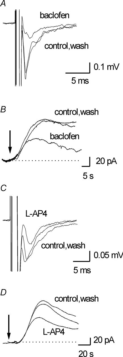
Adenosine is an important signalling molecule involved in a large number of physiological functions. In the brain these processes are as diverse as sleep, memory, locomotion and neuroprotection during episodes of ischaemia and hypoxia. Although the actions of adenosine, through cell surface G-protein-coupled receptors, are well characterized, in many cases the sources of adenosine and mechanisms of release have not been defined. Here we demonstrate the activity-dependent release of adenosine in the cerebellum using a combination of electrophysiology and biosensors. Short trains of electrical stimuli delivered to the molecular layer in vitro, release adenosine via a process that is both TTX and Ca(2+) sensitive. As ATP release cannot be detected, adenosine must either be released directly or rapidly produced by highly localized and efficient extracellular ATP breakdown. Since adenosine release can be modulated by receptors that act on parallel fibre-Purkinje cell synapses, we suggest that the parallel fibres release adenosine. This activity-dependent adenosine release exerts feedback inhibition of parallel fibre-Purkinje cell transmission. Spike-mediated adenosine release from parallel fibres will thus powerfully regulate cerebellar circuit output.


Figures

Figure 1
Figure 1. Electrical stimulation in the molecular layer releases adenosine
A, superimposed current traces from adenosine (Ado) and null biosensors following electrical stimulation of the molecular layer (5 V for 5 s at arrow). The lack of signal on the null sensor indicates that the current on the adenosine biosensor is due to purine detection and is not non-specific. B, application of 10 μm adenosine (Ado) and then 10 μm inosine (Ino) produced current responses on an adenosine biosensor. Addition of 20 μm EHNA (to block adenosine deaminase) almost abolished the response to adenosine but had little effect on the response to inosine. C, superimposed traces from an adenosine biosensor in control and in the presence of 20 μm EHNA (to block adenosine deaminase) following electrical stimulation of the molecular layer (8 V for 8 s at arrow). EHNA reduced the current by ∼75% demonstrating that most of the signal is due to adenosine detection. All stimulations were at a frequency of 20 Hz.
Figure 2
Figure 2. Properties of adenosine release
A, graph summarizing how adenosine release varies with stimulation frequency (data from 4 slices). The number of stimuli was kept constant (100) and the stimulation frequency was varied between 2 and 80 Hz. The data are normalized to the concentration of adenosine detected at a stimulus frequency of 20 Hz. B, superimposed traces from an adenosine biosensor following 20, 100 and 160 stimuli. The stimulus frequency was kept constant at 20 Hz. The stimulation strength was between 3 and 8 V (A) and 5 V (B).
Figure 3
Figure 3. Mechanisms of adenosine release
A, superimposed current traces from an adenosine biosensor placed on the surface of the molecular layer in control, in 10 μm CNQX + 50 μm AP5 and in 0.5 μm TTX. The adenosine release following electrical stimulation (5 V, 8 s at arrow) was abolished by TTX but was insensitive to the block of glutamate receptors. B, superimposed traces from an adenosine biosensor placed on the surface of the molecular layer in control, Ca2+-free aCSF (3.7 mm Mg2+) and following reintroduction of Ca2+ (wash). The adenosine release following electrical stimulation (7 V, 8 s at arrow) was abolished by removal of Ca2+. Inset, graph plotting normalized adenosine release against external Ca2+ concentration. Adenosine release was normalized to what occurred at 2.4 mm Ca2+ and summarizes data from 6 slices. C, superimposed traces from an adenosine biosensor placed on the surface of the molecular layer in control and in the presence of the GABAA receptor agonist muscimol (30 μm). Adenosine was released by a 5 V, 7 s stimulus at the arrow. Inset, superimposed averages of EPSPs in control and in 30 μm muscimol (*). Muscimol reduced EPSP amplitude but had little effect on the volley. D, superimposed traces from an adenosine biosensor placed on the surface of the molecular layer in control and in the presence of 5 μm NBTI and 10 μm dipyridamole to block equilibrative transport. Adenosine was released by a 5 V, 7 s stimulus at the arrow. All stimuli were at a frequency of 20 Hz.
Figure 4
Figure 4. ATP release is not detected
A, superimposed traces from an adenosine biosensor placed on the surface of the molecular layer and an ATP biosensor placed within the molecular layer. To maximize the likelihood of detecting ATP, adenosine was first detected (by an adenosine biosensor) then the ATP biosensor was placed in the molecular layer where the adenosine was detected. Following stimulation (arrow, 10 s) although adenosine was detected, no ATP release was measured. B, superimposed traces from an adenosine biosensor placed on the surface of the molecular layer and an ATP biosensor placed within the molecular layer. Following the application of the ecto-ATPase inhibitor Evans Blue (100 μm) the adenosine current was not diminished and there was still no ATP detection. The traces in B are from the same slice as in A. C, superimposed traces from an adenosine biosensor placed on the surface of the molecular layer and an ATP biosensor placed within the layer as in A and B. The biosensors were positioned at an approximately equal distance from the stimulating electrode. TTX (0.5 μm) was present to block TTX-dependent adenosine release. Stimulation (30 V, 10 s) caused cell damage and resulted in the electroporation of ATP, which produced a large current on the ATP biosensor. A small proportion of the released ATP (∼20%) was broken down to adenosine and was detected by the adenosine biosensor. The traces in C have been scaled by sensor calibration. All stimulations were at 20 Hz.
Figure 5
Figure 5. Metabolism of ɛ-ATP by cerebellar slices at room temperature
A, graph plotting percentage composition against time for the breakdown of 50 μmɛ-ATP. At time zero there was ∼90% ATP (▪) and 10% ADP (▴). Following incubation there was a fall in the proportion of ATP and a subsequent increase in AMP (□) and adenosine (^) with little change in ADP. B, graph plotting percentage of 50 μmɛ-ATP remaining against time in control and in the presence of inhibitors (Evans Blue and ARL67156). C, graph plotting percentage of adenosine against time in control and in the presence of inhibitors during metabolism of 50 μmɛ-ATP. α,β-Methylene-ADP (100 μm) was the most effective in slowing the build-up of adenosine. D, graph plotting percentage composition against time for the breakdown of 5 μmɛ-ATP. There was a more rapid breakdown of 5 μm ATP compared with 50 μmɛ-ATP (compare with A). E, graph plotting percentage of ɛ-ATP (5 μm) remaining against time in control and in the presence of inhibitors. Note that both Evans Blue and ARL67156 (100 μm) were more effective at slowing the metabolism of 5 μmversus 50 μmɛ-ATP (compare E and B). F, graph plotting percentage of adenosine against time in control and in the presence of inhibitors during metabolism of 5 μmɛ-ATP. Again inhibitors are more effective at slowing build up of adenosine with lower concentrations of substrate. Graphs A, B and C summarize data from 4 experiments; graphs D, E and F are from 3 experiments.
Figure 6
Figure 6. Adenosine release is modulated by parallel fibre receptor agonists
Parallel fibre (PF) EPSPs were evoked every 10 s by a stimulating electrode placed on the surface of the molecular layer and recorded with an extracellular electrode. A, superimposed averages of EPSPs in control, in 25 μm baclofen and following wash. Baclofen reversibly reduced EPSP amplitude by ∼60%. B, superimposed traces from an adenosine biosensor in control, baclofen (25 μm) and in wash. Adenosine release was evoked by a 10 s stimulus at the arrow. Baclofen reversibly reduced adenosine release by ∼50%. C, superimposed averages of EPSPs in control, in 50 μm l-AP4 and following wash. l-AP4 reversibly reduced EPSP amplitude by ∼40%. D, superimposed traces from an adenosine biosensor placed on the surface of the molecular layer in control, 50 μm l-AP4 and in wash. Following l-AP4 application adenosine release was decreased by ∼40%. Adenosine release was evoked by a 5 s stimulus (20 Hz) at the arrow.
Figure 7
Figure 7. The adenosine released by electrical stimulation modulates parallel fibre–Purkinje cell synaptic transmission
Parallel fibre (PF) EPSPs were evoked every 10 s by a stimulating electrode placed on the surface of the molecular layer and recorded with an extracellular electrode. A, graph plotting PF EPSP amplitude against time. Application of 1 μm 8CPT, to block A1 receptors, increased EPSP amplitude by ∼30%. B, superimposed traces from an adenosine biosensor in control and in the presence of 1 μm 8CPT. Adenosine release was evoked by a 7 s stimulus at the arrow. Application of 8CPT increased adenosine release by 43%. C, graph plotting PF EPSP (evoked every 10 s) amplitude against time. At the asterisk a train of stimuli was delivered (10 s, 20 Hz) to cause adenosine release. Following the train there was a marked reduction in PF EPSP amplitude followed by a slow recovery back to control amplitude (PF EPSP amplitude during the train is not plotted). The time course of PF EPSP amplitude recovery was ∼150 s which is very similar to the time course of adenosine release. Application of the A1 receptor antagonist 8CPT increased PF EPSP amplitude by ∼25% and also markedly speeded recovery following a train of stimuli (∼50 s). Thus, the slow component of recovery results from the released adenosine activating A1 receptors. D, graph summarizing data from 6 recordings. Following blockade of A1 receptors there was significant speeding of EPSP recovery.

Similar articles

Cited by

References

    1. Agarwal RP, Spector T, Parks RE. Tight-binding inhibitors-IV. Inhibition of adenosine deaminases by various inhibitors. Biochem Pharmac. 1977;26:359–367. - PubMed
    1. Batchelor AM, Garthwaite J. GABAB receptors in the parallel fibre pathway of rat cerebellum. Eur J Neurosci. 1992;4:1059–1064. - PubMed
    1. Beierlein M, Regehr WG. Brief bursts of parallel fiber activity trigger calcium signals in Bergmann glia. J Neurosci. 2006;26:6958–6967. - PMC - PubMed
    1. Billups B, Graham BP, Wong AYC, Forsythe ID. Unmasking group III metabotropic glutamate autoreceptor function at excitatory synapses in the rat CNS. J Physiol. 2005;565:885–896. - PMC - PubMed
    1. Braas KM, Newby AC, Wilson VS, Snyder SH. Adenosine-containing neurons in the brain localized by immunocytochemistry. J Neurosci. 1986;6:1952–1961. - PMC - PubMed

Publication types

MeSH terms