. 2007 Jul;37(1):97-104.
doi: 10.1165/rcmb.2006-0419OC. Epub 2007 Mar 8.

Measurement of IL-13-induced iNOS-derived gas phase nitric oxide in human bronchial epithelial cells

Affiliations

Measurement of IL-13-induced iNOS-derived gas phase nitric oxide in human bronchial epithelial cells

Vinod Suresh et al. Am J Respir Cell Mol Biol. 2007 Jul.

Abstract

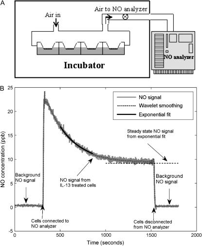
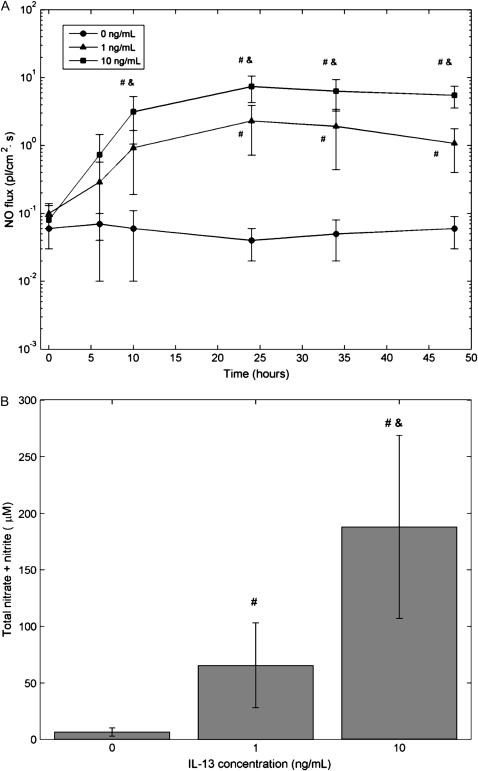
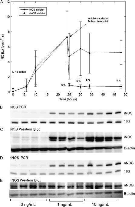
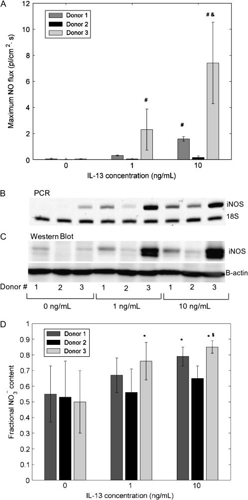
Exhaled nitric oxide (NO) is altered in numerous diseases including asthma, and is thought broadly to be a noninvasive marker of inflammation. However, the precise source of exhaled NO has yet to be identified, and the interpretation is further hampered by significant inter-subject variation. Using fully differentiated normal human bronchial epithelial (NHBE) cells, we sought to determine (1) the rate of NO release (flux, pl.s(-1.)cm(-2)) into the gas; (2) the effect of IL-13, a prominent mediator of allergic inflammation, on NO release; and (3) inter-subject/donor variability in NO release. NHBE cells from three different donors were cultured at an air-liquid interface and stimulated with different concentrations of IL-13 (0, 1, and 10 ng/ml) for 48 h. Gas phase NO concentrations in the headspace over the cells were measured using a chemiluminescence analyzer. The basal NO flux from the three donors (0.05 +/- 0.03) is similar in magnitude to that estimated from exhaled NO concentrations, and was significantly increased by IL-13 in a donor-specific fashion. The increase in NO release was strongly correlated with inducible nitric oxide synthase (iNOS) gene and protein expression. There was a trend toward enhanced production of nitrate relative to nitrite as an end product of NO metabolism in IL-13-stimulated cells. NO release from airway epithelial cells can be directly measured. The rate of release in response to IL-13 is strongly dependent on the individual donor, but is primarily due to the expression of iNOS.


Figures

<b>Figure 1.</b>
Figure 1.
Real-time measurement of gas phase NO release by NHBEs (A) Schematic of gas phase NO measurement apparatus. The lids of 12-well Transwell plates were modified to form a gas tight seal. The headspace of the Transwells containing epithelial monolayers was perfused with air at a constant flow rate. The effluent air was fed to an NO analyzer to measure NO concentrations. (B) One representative measurement of the real-time response of NO analyzer shows a low background level in the incubator air. Switching the analyzer intake to effluent air from the headspace over the cells causes a sharp spike in the response that gradually decays to a plateau. Switching the intake back to the incubator air results in a low background NO reading. The raw analyzer response (solid gray line) is smoothed using a wavelet transform (dotted gray line). The steady-state NO concentration at the plateau is determined by fitting an exponential equation to 300 data points of the smoothed curve.
<b>Figure 2.</b>
Figure 2.
IL-13 increases gas phase NO and total nitrate content in culture medium. (A) Different concentrations of IL-13 (0, 1, 10 ng/ml) were introduced in the culture medium of NHBEs at time t = 0. Gas phase NO release was followed over a period of 48 h by determining steady-state NO concentrations at different times and calculating a flux as described in Materials and Methods. Basal (0 ng/ml) NO flux was small and relatively constant. IL-13 caused a dose-dependent increase in NO flux by up to 2 orders of magnitude (n = 4 or 12; #,&: P < 0.05 compared with 0 and 1 ng/ml, respectively). Results for donor 3 cells; other donors showed similar trends with smaller magnitudes. (B) Total nitrate content in the culture medium shows a dose-dependent increase after the cells were exposed to IL-13 for 48 h (n = 13 or 14) (#,&: P < 10−5 compared with 0 and 1 ng/ml, respectively).
<b>Figure 3.</b>
Figure 3.
IL-13–mediated increase in NO flux shows a significant decrease with iNOS, but not nNOS, inhibitor and correlates with iNOS mRNA and protein expression. (A) IL-13 (10 ng/ml) was added to the culture medium at time t = 0 (2 groups, n = 4 each) and an increase in NO flux observed for the first 24 h. At t = 24 h, an iNOS or nNOS competitive inhibitor was added to the culture medium of each group. The iNOS inhibitor reduced NO flux by more than 85% within 1 h of addition, while the nNOS inhibitor reduced NO flux by < 30% ($: P < 0.01 compared with flux at 24 h, %: P < 0.05 compared with nNOS inhibitor). IL-13 upregulates iNOS gene (B) and protein expression (C). nNOS gene (D), but not protein (E) expression is upregulated at an IL-13 concentration of 1 ng/ml. RT-PCR products and Western blot show replicates from four experiments using donor 3 cells; other donors showed similar trends.
<b>Figure 4.</b>
Figure 4.
Significant donor variation in NO flux and relative nitrate/nitrite content in culture medium correlates with iNOS gene and protein expression. Cells from different donors show significantly different response to IL-13 as measured by the maximum NO flux after exposure (A), iNOS gene (B), and protein (C) expression. Donor 3 cells are most responsive to increasing IL-13 concentrations, while donor 2 cells are least responsive (n = 6 or 12; #: P < 0.001 compared with donor 2, &: P < 0.01 compared with donor 1). Individual amounts of nitrite formula image) and nitrate formula image) in the culture medium were determined after 48 h exposure to IL-13, and the fractional nitrate content [ formula image / formula image + formula image) ] was calculated. Fractional formula image content was close to 0.5 for all donors under basal conditions (0 ng/ml) and increased concomitant with the IL-13–induced upregulation in iNOS mRNA and protein (D) (n = 6, 13, or 14; *: P < 0.02 compared with 0 ng/ml, $: P < 0.0001 compared with 1 ng/ml).

References

    1. Barnes P, Kharitonov S. Exhaled nitric oxide: a new lung function test. Thorax 1996;51:233–237. - PMC - PubMed
    1. Smith AD, Cowan JO, Brassett KP, Herbison GP, Taylor DR. Use of exhaled nitric oxide measurements to guide treatment in chronic asthma. N Engl J Med 2005;352:2163–2173. - PubMed
    1. Moodley YP, Lalloo UG. Exhaled nitric oxide is elevated in patients with progressive systemic sclerosis without interstitial lung disease. Chest 2001;119:1449–1454. - PubMed
    1. Rolla G, Colagrande P, Brussino L, Bucca C, Bertero M, Caligaris-Cappio F. Exhaled nitric oxide and pulmonary response to iloprost in systemic sclerosis with pulmonary hypertension. Lancet 1998;351:1491–1492. - PubMed
    1. Buchvald F, Baraldi E, Carraro S, Gaston B, De Jongste J, Pijnenburg MW, Silkoff PE, Bisgaard H. Measurements of exhaled nitric oxide in healthy subjects age 4 to 17 years. J Allergy Clin Immunol 2005;115:1130–1136. - PubMed

Publication types