Review
. 2007 Mar;71(1):158-229.
doi: 10.1128/MMBR.00036-06.

Colicin biology

Affiliations
Review

Colicin biology

Eric Cascales et al. Microbiol Mol Biol Rev. 2007 Mar.

Abstract

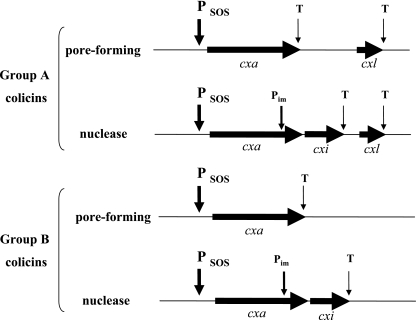
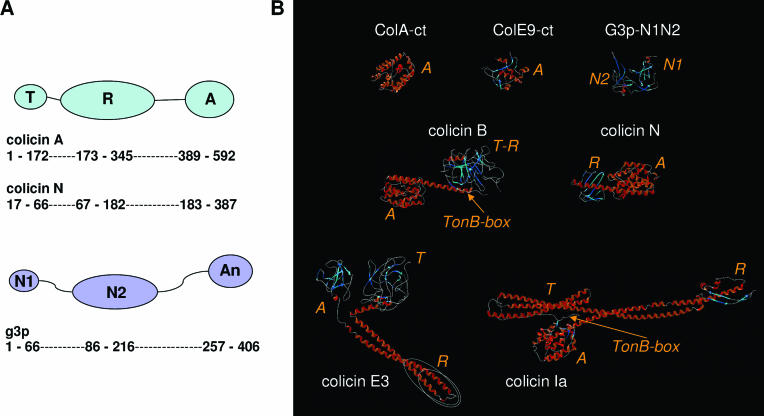
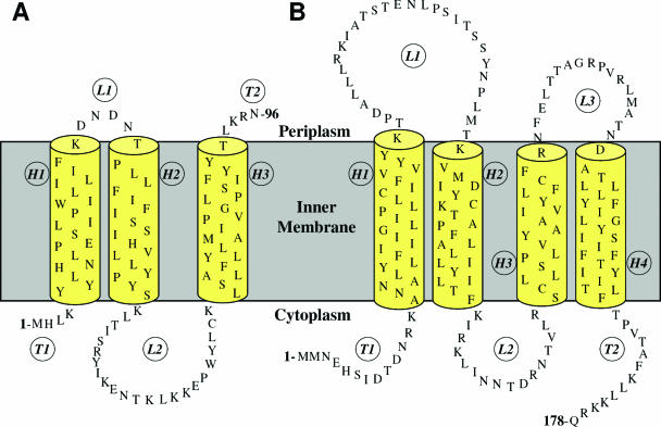
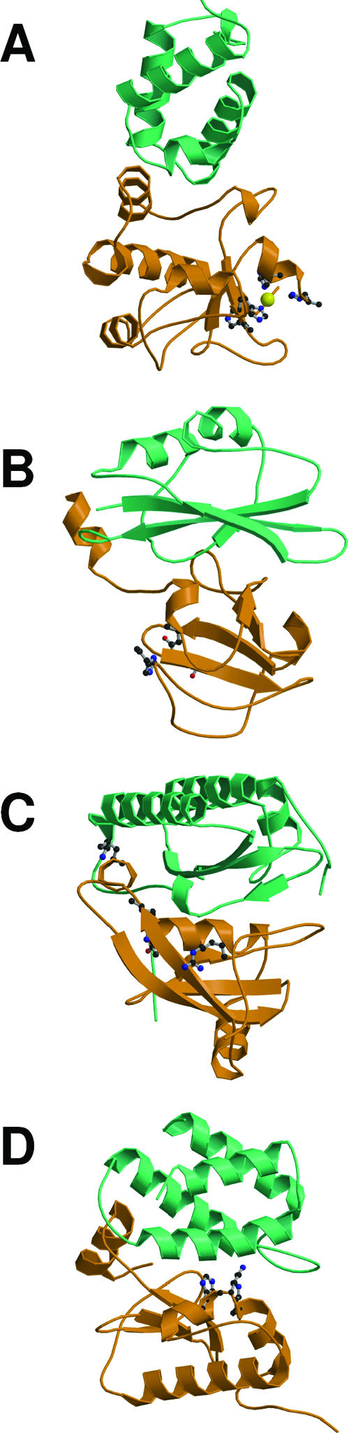
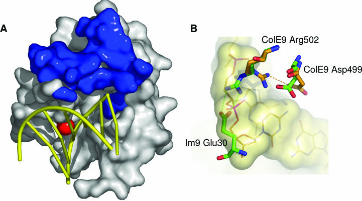
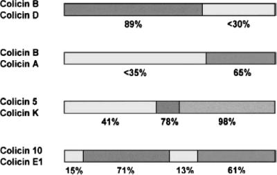
Colicins are proteins produced by and toxic for some strains of Escherichia coli. They are produced by strains of E. coli carrying a colicinogenic plasmid that bears the genetic determinants for colicin synthesis, immunity, and release. Insights gained into each fundamental aspect of their biology are presented: their synthesis, which is under SOS regulation; their release into the extracellular medium, which involves the colicin lysis protein; and their uptake mechanisms and modes of action. Colicins are organized into three domains, each one involved in a different step of the process of killing sensitive bacteria. The structures of some colicins are known at the atomic level and are discussed. Colicins exert their lethal action by first binding to specific receptors, which are outer membrane proteins used for the entry of specific nutrients. They are then translocated through the outer membrane and transit through the periplasm by either the Tol or the TonB system. The components of each system are known, and their implication in the functioning of the system is described. Colicins then reach their lethal target and act either by forming a voltage-dependent channel into the inner membrane or by using their endonuclease activity on DNA, rRNA, or tRNA. The mechanisms of inhibition by specific and cognate immunity proteins are presented. Finally, the use of colicins as laboratory or biotechnological tools and their mode of evolution are discussed.


Figures

FIG. 1.
FIG. 1.
Organization of the colicin operons. The genes are represented by arrowheads. SOS promoters (PSOS), the immunity promoter (Pim), and transcription terminators (T) are indicated by arrows. Names of the colicin gene (cxa, in which x is specific to the colicin) and its immunity gene (cxi) and lysis protein gene (cxl) follow the nomenclature.
FIG. 2.
FIG. 2.
Sequence alignment of colicin lysis proteins. Amino acid sequences of the colicin lysis proteins are shown. The sequences of the colicin lysis proteins encoded by the colicinogenic plasmids indicated at the left are presented. Identical amino acids are in boldface type. The numbers of residues in the signal peptide and in the mature form are indicated. X63621, EMBL/GenBank/DDBJ accession number X63621.
FIG. 3.
FIG. 3.
Schematic summary of reception, translocation, and mode of action of most studied colicins. Colicins are distinguished by their general modes of action (upper section, enzymatic; lower section, pore forming) and transit machineries (right section, TonB; left section, Tol) separated by dotted lines. For each colicin (the name is indicated at the arrow base), the outer membrane protein used for the reception step (and sometimes for outer membrane translocation) (ColB, D, Ia, Ib, M, and N) and the OM protein involved in the translocation step (OmpF, colicins A, E2 to E9, K, and U; TolC, colicins 5, 10, and E1) are indicated. For enzymatic colicins, the mode of action at the physiology level (peptidoglycan synthesis block [colicin M], protein synthesis block by cleavage of tRNA [colicins D and E5] or 16S rRNA [colicins E3, E4, and E6], and DNA degradation [colicins E2 and E7 to E9]) is also indicated. For tRNA colicins (colicins D and E5), the specific tRNA targeted is indicated by the one-letter code in parentheses (D, aspartate; H, histidine; N, asparagine; R, arginine; Y, tyrosine). See the text for details.
FIG. 4.
FIG. 4.
Colicin and g3p domain organization and crystal structures. (A) Schematic representation of the domains involved in reception (R, N2), translocation (T, N1), and activity (A) (for colicins) or anchoring (An) (for g3p). Positions of residues surrounding the various domains are indicated together with hinge regions connecting the domains. Colicin A, N, and g3p domains are presented. (B) Structures of the C-terminal domains of colicin A (ColA-ct) (PDB accession number 1COL; resolution, 2.40 Å) and colicin E9 (ColE9-ct) (PDB accession number 1EMV; resolution, 1.70 Å) and the structures of intact colicin E3 (PDB accession number 1JCH; resolution, 3.02 Å), colicin Ia (PDB accession number 1CII; resolution, 3.00 Å), colicin B (PDB accession number 1RH1; resolution, 2.50 Å), colicin N (PDB accession number 1A87; resolution, 3.10 Å), and g3p (reception-N2 and translocation-N1 domains) (PDB accession number 1G3P; resolution, 1.46 Å). The Im9 and Im3 proteins bound to colicin E9 and colicin E3, respectively, have been removed from the complex structures. The TonB box sequences present in the first β-strand of colicin B and in the first residues of colicin Ia are indicated. The receptor binding domains of colicin E3 and E9 (76 residues from colicin E9) (512) or corresponding to a cyclic peptide of 34 residues from colicin E3 (463) are indicated by circles on the colicin E3 three-dimensional structure.
FIG. 5.
FIG. 5.
Ribbon diagrams of BtuB-colicin E3 and Cir-colicin Ia. (A) BtuB-colicin E3 complex (371) (PDB accession number 1UJW; 2.75 Å). BtuB and part of the receptor binding domain of colicin E3 are colored dark gold, while the plug domain of BtuB is colored light gold. Several β-strands have been removed to show the plug domain more clearly. Colicin E3 is seen at the top of the figure as a long coiled coil with a loop between helices that interacts with extracellular loops of BtuB. Residues of this loop that participate in receptor recognition are shown in the space-filling representation. (B) Cir-colicin Ia complex (Buchanan et al., unpublished). Cir and colicin Ia are shown in dark blue, with light blue representing the plug domain. Again, several β-strands have been removed to visualize the plug domain. The receptor binding domain of colicin Ia (at the top of the figure) consists of two β-strands wrapped around a long α-helix, and the space-filling representation depicts colicin Ia residues that interact with Cir. Dotted lines indicate residues not seen in the crystal structure due to high mobility (disorder). (C) A superposition of the two complex structures shows that the tips of colicin E3 and colicin Ia extend to about the same depth in the respective transporters. Each colicin binds its transporter at an angle of approximately 45° with respect to the lipid bilayer although from opposite directions. Courtesy of Petra Lukacik, reproduced with permission.
FIG. 6.
FIG. 6.
Localization and topologies of components of colicin import machineries. (A) Topologies and predicted transmembrane domains of the inner membrane TolA, TolQ, and TolR proteins. The periplasmic TolB and the outer membrane-anchored peptidoglycan-associated lipoprotein Pal are shown. The Pal peptidoglycan binding sequence and TolQRA transmembrane segment boundaries are indicated (39, 78, 119, 400, 472, 651). (B) Topologies and predicted transmembrane domains of TonB, ExbB, and ExbD. Two predictions for ExbB transmembrane domains have been made (320, 325). Those described by Karlsson et al. (325) are in parentheses. ext, external medium; peri, periplasm; cyto, cytoplasm.
FIG. 7.
FIG. 7.
TolA, TolB, and Pal crystal structures. (A) Ribbon diagrams of TolA, TolB, and Pal. The Pseudomonas aeruginosa TolA C-terminal domain (TolAIII) (677) (PDB accession number 1LRO; 1.80 Å) is shown. The α-helix indicated by the arrow corresponds to the C-terminal 43 residues of the central domain (TolAII). (B) The Escherichia coli TolB structure (1, 72) (PDB accession number 1CRZ; 1.95 Å) showing the D1 domain and the six-bladed D2 domain (β-propeller) forming the cavity. (C) The Escherichia coli Pal lipoprotein structure (C. Abergel and A. Walburger, unpublished data) (PDB accession number 10AP; 1.93 Å). The peptidoglycan binding helix is indicated with an arrow, whereas the lateral chain of the Tyr residue of the conserved TolA binding motif (SYGK) (Fig. 8B) is shown and highlighted by the pink circle.
FIG. 8.
FIG. 8.
Tol binding sequences. (A) Schematic illustration of the colicin A N-terminal domain and the three binding sequences identified (RBS, TolR-binding sequence, residues 7 to 11; BBS, TolB-binding sequence, residues 7 to 20; ABS, TolA-binding sequence, residues 52 to 97) (42, 43, 313). (B) Sequence alignments of the Pal, colicins A, K, and N, and g3p TolA binding sequences. Conserved residues are in boldface type, and the TolA binding motif with the conserved and critical tyrosine residue is highlighted in green (78, 527). Lower panel, results from alanine-scanning mutagenesis of residues 56 to 75 of colicin N (228). Uppercase letters, essential residues; lowercase letters, nonessential residues. (C) Sequence alignments of the TolB-dependent (upper panel) and -independent (lower panel) colicin TolB binding sequences. Conserved residues are in boldface type. Colicin TolB boxes are highlighted in blue, whereas the colicin A TolR binding sequence is framed. Note that the WSSE motif present in TolB-dependent colicin TolB binding sequences is not found in TolB-independent colicins. Middle panel, results from alanine-scanning mutagenesis of residues 34 to 44 of colicin E9 (248). Large letters, critical residues; small letters, nonessential residues; medium letters, mutations decreasing but not abolishing TolB-colicin E9 N-terminal domain affinity. (D) Crystal structure of the TolB protein (green) with the residues 32 to 47 of colicin E9 (purple) (417) showing localization of the TolB binding sequence at the entrance of the β-propeller. (E) Top view of the same complex, which emphasizes the binding site. The colicin E9 peptide is represented in a backbone. (Panels D and E are reprinted from reference with permission of the publisher. Copyright 2006 National Academy of Sciences U.S.A.)
FIG. 9.
FIG. 9.
Model for colicin B transport across the outer membrane. In step 1, binding of colicin B (blue) to FepA (red) causes the FepA TonB box (A) to become periplasmically exposed. In step 2, TonB releases FepA globular domain bound to colicin B. The colicin B TonB box (B) can now bind TonB. In step 3, TonB releases colicin B into the periplasm. TonB is not shown on this model because its active configuration at the outer membrane is not certain. (Reprinted from reference with permission from Blackwell Publishing.)
FIG. 10.
FIG. 10.
BtuB-TonB and FhuA-TonB crystal structures. (A) BtuB-TonB view from the periplasm. The BtuB barrel is in orange, vitamin B12 is shown as red spheres, the BtuB internal globular domain is in green, and the TonB carboxy terminus (amino acids 153 to 233) is in magenta, with the TonB box highlighted in blue. (B) BtuB-TonB side view from within the outer membrane. (Panels A and B are reprinted from reference with permission of AAAS.) (C) FhuA-TonB side view from within the outer membrane. The FhuA barrel is in blue, the FhuA internal globular domain is in green, and the TonB carboxy terminus (amino acids 158 to 235) is in yellow. (D) FhuA-TonB view from the periplasm. TonB α-helices (α1 and α2) and β-strands are labeled, as are periplasmic turns in FhuA (T1 and T7 to T10), for reference. (Panels C and D are reprinted from reference with permission of AAAS.)
FIG. 11.
FIG. 11.
The TonB box. (A) Sequence alignments of TonB boxes. Shown are sequence alignments of the BtuB, Cir, FecA, FepA, and FhuA (upper panel) and of the colicin B, D, Ia/Ib, M, and 5/10 and pesticin TonB boxes (lower panel). Conserved residues are in boldface type. (B) BtuB TonB box interactions with TonB. Shown are in vivo cysteine disulfide cross-linking data for the TonB region around Gln160 (green) and the BtuB TonB box (blue). The antiparallel interaction between the β-strands is shown, as observed in the crystal structure. (Reprinted from reference with permission of AAAS.) Solid lines indicate cross-links observed between TonB and BtuB (67). Dotted lines indicate cross-links observed between TonB and FecA, with the FecA TonB box aligned with the BtuB TonB box (492).
FIG. 12.
FIG. 12.
Space-filling model of the TonB NMR monomer (509). Backbone atoms are gray, aromatic side chains (F180, F202, W213, Y215, and F230) are yellow, and other side chains are magenta.
FIG. 13.
FIG. 13.
Speculative models of colicin import. (A) Import of group A colicins. In stage 1, the colicin contacts the bacterial cell and binds to the outer membrane receptor by its central domain. In stage 2, the colicin partly unfolds and recruits the translocation machinery (e.g., the outer membrane translocon and the Tol complex). In stage 3, the colicin N-terminal domain translocates through the OM β-barrel and protrudes into the periplasm, where it interacts with the TolB subunit, thus dissociating the TolB-Pal complex. Stages 4 and 5 depict the Brownian ratchet mechanism. The colicin N-terminal domain dissociates from TolB to interact with TolA (stage 4) and then with TolQ and/or TolR (stage 5), probably after TolA degradation. In stage 6, the colicin C-terminal domain, carrying the lethal activity, is translocated by an unknown mechanism (?) and forms a pore in the inner membrane or translocates to the cytoplasm to degrade nucleic acids. The extracellular face is on the top, and the cytoplasm is on the bottom. A, TolA; A*, degradation product of TolA; B, TolB; P, Pal; Q, TolQ; R, TolR; Rec, OM receptor; T, OM translocon. Colicin is in red (t, translocation domain; r, reception domain; a, activity domain). (B) Import of group B colicins. In stage 1, the colicin contacts the bacterial cell and binds to a TonB-dependent gated channel by its central domain. In stage 2, the receptor recruits the TonB machinery through the colicin-induced receptor TonB box accessibility. In stage 3, following energy input by the ExbBD-TonB complex (yellow flash), the colicin N-terminal domain translocates through the OM and interacts with TonB by its own TonB box. Please note that two models are currently envisaged. The second model implies that TonB shuttles from the IM to the OM to release stored energy (see the group B colicin transit section). In stage 4, the colicin N-terminal domain dissociates from TonB to interact with ExbB and/or ExbD. In stage 5, the colicin C-terminal domain, carrying the lethal activity, is translocated by an unknown mechanism (?) to reach its final compartment. The extracellular face is on the top, and the cytoplasm is on bottom. B, ExbB; D, ExbD; Rec, TonB-dependent OM-gated channel. Colicin is in red (t, translocation domain; r, reception domain; a, activity domain). The TonB boxes of the receptor and the colicin are indicated by trapezoids. Another speculative model for group B colicin translocation is depicted Fig. 9.
FIG. 14.
FIG. 14.
Crystal structure of g3p-N1 bound to TolAIII. The cocrystal structure of the M13 g3p translocation domain (g3p-N1, left) with the TolA C-terminal domain (TolAIII, right) (PDB accession number 1TOL; 1.85 Å) (421) is shown. The conserved TolA binding motif (CYGT) (Fig. 8B) of g3p-N1, located at the interface between the two domains, is shown in a ball-and-stick representation and is highlighted by a pink circle.
FIG. 15.
FIG. 15.
Speculative model for Tol-dependent filamentous phage DNA translocation. The model is depicted in six consecutive stages. In stage 1, the filamentous bacteriophage contacts the bacterial cell and binds to the tip of an F pilus by its reception N2 domain (r). In stage 2, the interaction between g3p-N2 and the F pilus dissociates g3p-N1. In stage 3, the F pilus retracts and brings the g3p molecule into the periplasm, where it interacts through the translocation domain (t) with the TolA C-terminal domain (TolAIII). In stage 4, the g3p-N2 domain interacts with the TolA central domain (TolAII). In stage 5, the g3p-N3 domain that anchors the minor coat protein into the phage particle dissociates, inserts into the inner membrane, oligomerizes, and forms pores by which the single-stranded DNA (in red) translocates (stage 6) after disaggregation of the capsid by the insertion of the major coat g8p proteins into the membrane. The TolA degradation product observed upon the treatment of WT cells with M13 bacteriophage is represented (A*). Periplasm is on the top, and cytoplasm is on the bottom. The insert at stage 2 represents the electron microscopy picture of an M13 filamentous bacteriophage bound at an F pilus tip (bar, 100 nm). (Reprinted from reference with permission.)
FIG. 16.
FIG. 16.
Comparison of the structures of the pore-forming domains of colicins A, N, B, Ia, and E1. Each molecule is shown in two orientations: one approximately parallel to the hydrophobic hairpin (upper) and the other perpendicular to it (lower). The hydrophobic helices are red, whereas the other eight helices are blue. Helix numbers are shown explicitly for colicin A. The overall similarity in the folds of these molecules is apparent.
FIG. 17.
FIG. 17.
Schematic representation of certain closed states. Closed colicin channels can adopt several membrane-bound conformations, two of which are illustrated. (A) Penknife model based on studies of colicin A (374). (B) Variant of the umbrella model, as proposed for colicin E1 (408). The figure is not meant to suggest that the secondary structure of the protein in these states replicates the crystal structure.
FIG. 18.
FIG. 18.
Schematic representations of the transmembrane segments of the open colicin channel. Only the channel-forming domain is shown. The depicted arrangement of the protein segments within the membrane derives from experiments, but the secondary structure (shown as blue, numbered, α-helical cylinders) follows that of the crystal structure and is purely speculative. Likewise, the distortion of the lipid bilayer is shown only as an acknowledgment of the potential involvement of lipids in the structure and not as a specific model. (A) Colicin Ia. Helices 2 to 5 are translocated across the membrane during channel opening. The inset shows the electrophysiological record of colicin Ia channels in a voltage-clamped lipid bilayer. Two channels open at positive voltage and close at negative voltage. (B) Colicin A. The colicin A channel resembles that of colicin Ia, but helices 2 and 5 are thought to be incompletely translocated. The inset shows colicin A channels in a lipid bilayer.
FIG. 19.
FIG. 19.
Schematic representation of the transmembrane segments of the open states of the isolated C-terminal domain of colicin Ia. (A) Normal conductance channel similar to the channel formed by the whole molecule. (B) Small conductance channel, the result of the translocation of H1. The inset shows the electrophysiological record showing the transition from the normal to the small conductance state. The membrane is clamped at +70 mV.
FIG. 20.
FIG. 20.
Topographical models of (A) Cfi and (B) Cai in the cytoplasmic membrane of Escherichia coli. H1, H2, H3, and H4 denote the trans-membrane α-helices; L1, L2, and L3 denote loops; and T1 and T2 correspond to the N- and C-terminal ends, respectively.
FIG. 21.
FIG. 21.
Structures of colicin nuclease domains bound to their cognate immunity proteins highlighting exosite (top) and active-site (bottom) binding by their neutralizing inhibitors. (A) ColE9 DNase-Im9 complex (PDB accession number 1BXI). (B) ColE3 rRNase-Im3 (PDB accession number 1E44). (C) ColE5 tRNase-Im5 (PDB accession number 2FHZ). (D) ColD tRNase-ImD (PDB accession number 1V74). Colicin nucleases are shown in orange, and their Im proteins are shown in cyan. Each structure identifies key active-site residues and, in the case of the DNase, the catalytic metal ion (see the text for details). Courtesy of Irina Grishkovskaya, reproduced with permission.
FIG. 22.
FIG. 22.
The Im9 binding site partially overlaps the DNA binding site on the colicin E9 DNase. (A) Crystal structure of ColE9 H551A (equivalent to His103 in the isolated E9 DNase) bound to 8-mer dsDNA showing the bound catalytic Mg2+ ion (red sphere) in the context of the Im9 exosite (blue shading) (PDB accession number 1V14). The enzyme binds to the minor groove of the DNA, causing it to widen and bend. (B) Structural overlay showing the steric clash of the salt bridge between ColE9 Arg502 and Im9 Glu30 with DNA (shown as a molecular surface). In the DNA-bound structure, ColE9 Arg502 swings out of the way to form a salt bridge with ColE9 Asp499. ColE9 Arg502 is equivalent to Arg54 in the isolated E9 DNase domain. Courtesy of Maria Maté, reproduced with permission.
FIG. 23.
FIG. 23.
Pairwise comparisons of pore-forming colicin protein sequences. Values below each comparison indicate the percent sequence identity for the region indicated. Colicins are not drawn to scale.
FIG. 24.
FIG. 24.
Graph indicating the average number of total nucleotide replacements between pairs of nuclease-type colicin gene clusters (colicin pairs E2/E9 and E3/E6). Most of the divergence between colicins occurs in the immunity region of the gene cluster (composed of the immunity gene and the immunity binding region of the colicin gene).
FIG. 25.
FIG. 25.
Survey of colicin production and resistance in E. coli. Over 400 strains were isolated from two populations of feral mice in Australia over a period of 7 months. The isolates were scored for colicin production and resistance. (a) Colicin production is abundant, with just under 50% of the strains producing eight distinct colicin types. col represents nonproducer strains. (b) The majority of isolates are resistant to most co-occurring colicins. (c) A small proportion of the population is sensitive to co-occurring colicins.

References

    1. Abergel, C., E. Bouveret, J. M. Claverie, K. Brown, A. Rigal, C. Lazdunski, and H. Bénédetti. 1999. Structure of the Escherichia coli TolB protein determined by MAD methods at 1.95 A resolution. Structure 7:1291-1300. - PubMed
    1. Akutsu, A., H. Masaki, and T. Ohta. 1989. Molecular structure and immunity specificity of colicin E6, an evolutionary intermediate between E-group colicins and cloacin DF13. J. Bacteriol. 171:6430-6436. - PMC - PubMed
    1. Altieri, M., J. L. Suit, M. L. J. Fan, and S. E. Luria. 1986. Expression of the cloned ColE1 kil gene in normal and Kilr Escherichia coli. J. Bacteriol. 168:648-654. - PMC - PubMed
    1. Amati, P. 1964. Vegetative multiplication of colicinogenic factors after induction in Escherichia coli. J. Mol. Biol. 78:239-246. - PubMed
    1. Anderluh, G., I. Gokce, and J. H. Lakey. 2004. A natively unfolded toxin domain uses its receptor as a folding template. J. Biol. Chem. 279:22002-22009. - PubMed

Publication types

LinkOut - more resources