. 2007 Apr;117(4):978-88.
doi: 10.1172/JCI29176. Epub 2007 Mar 8.

SPDEF regulates goblet cell hyperplasia in the airway epithelium

Affiliations

SPDEF regulates goblet cell hyperplasia in the airway epithelium

Kwon-Sik Park et al. J Clin Invest. 2007 Apr.

Abstract

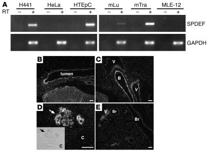
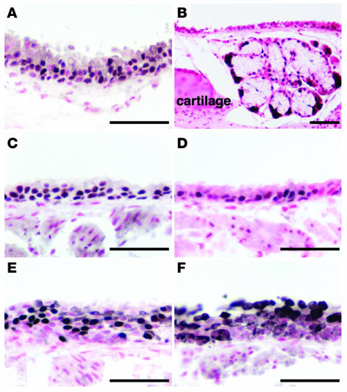
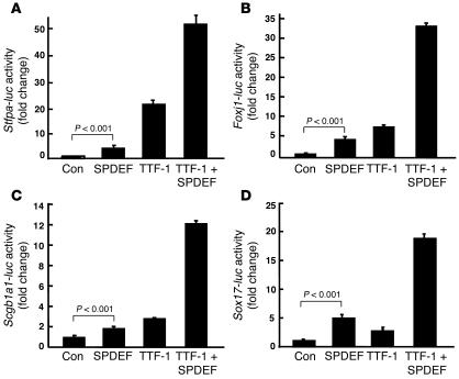
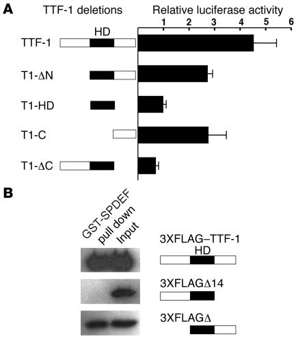
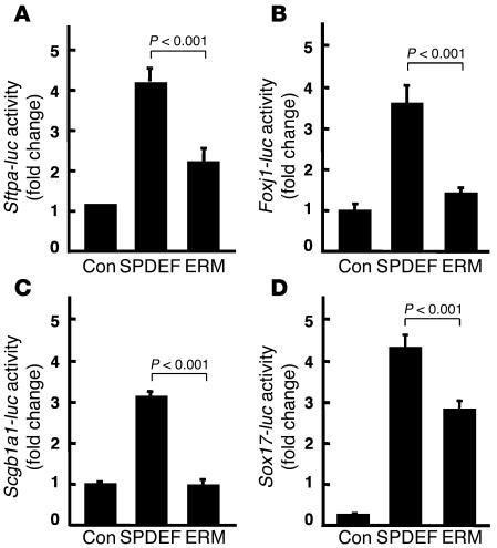
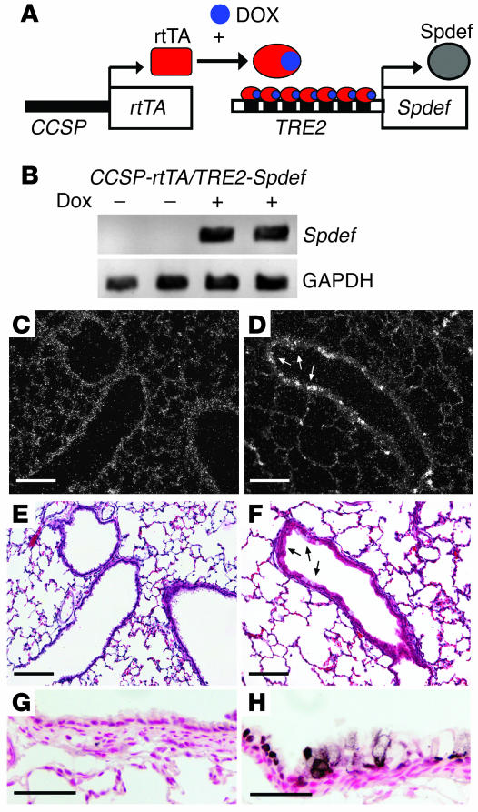
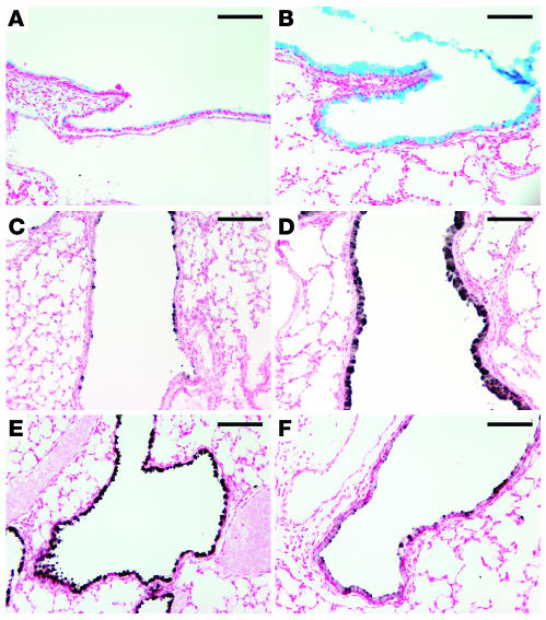
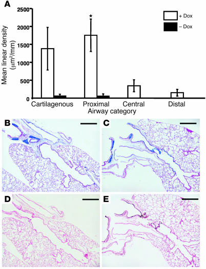
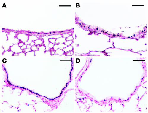
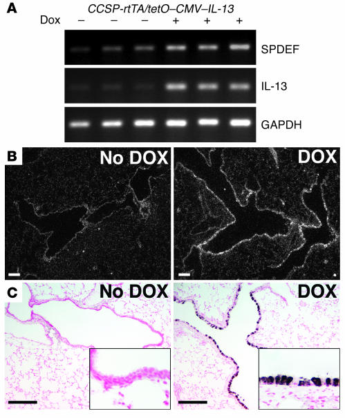
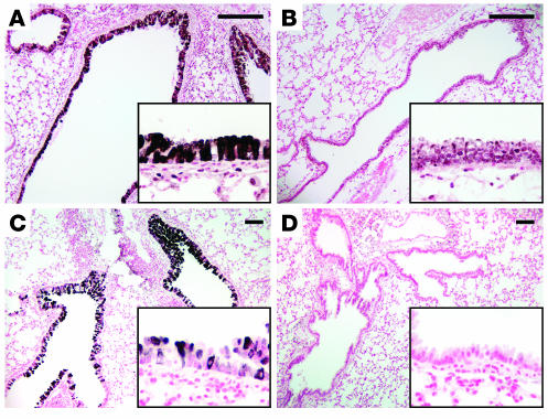
Goblet cell hyperplasia and mucous hypersecretion contribute to the pathogenesis of chronic pulmonary diseases including cystic fibrosis, asthma, and chronic obstructive pulmonary disease. In the present work, mouse SAM pointed domain-containing ETS transcription factor (SPDEF) mRNA and protein were detected in subsets of epithelial cells lining the trachea, bronchi, and tracheal glands. SPDEF interacted with the C-terminal domain of thyroid transcription factor 1, activating transcription of genes expressed selectively in airway epithelial cells, including Sftpa, Scgb1a1, Foxj1, and Sox17. Expression of Spdef in the respiratory epithelium of adult transgenic mice caused goblet cell hyperplasia, inducing both acidic and neutral mucins in vivo, and stainined for both acidic and neutral mucins in vivo. SPDEF expression was increased at sites of goblet cell hyperplasia caused by IL-13 and dust mite allergen in a process that was dependent upon STAT-6. SPDEF was induced following intratracheal allergen exposure and after Th2 cytokine stimulation and was sufficient to cause goblet cell differentiation of Clara cells in vivo.


Figures

Figure 1
Figure 1. Spdef mRNA in mouse respiratory epithelial cells, trachea, and tracheal glands.
(A) SPDEF and GAPDH expression was identified by RT-PCR using RNA extracts from human lung adenocarcinoma H441 cells, cervical adenocarcinoma HeLa cells, normal human tracheal epithelial HTEpC cells, SV40 large T immortalized mouse lung epithelial MLE-12 cells, mouse lung (mLu), and mouse trachea (mTra). Spdef mRNA was detected in H441 cells, HTEpC cells, and mouse lung and trachea, but not in HeLa or MLE-12 cells. PCR without RT (–) showed no product. (BE) In situ hybridization for Spdef mRNA was performed on sections of trachea and tracheal glands (B and D) and lung (C and E) in adult mice. Spdef mRNA was detected in the epithelium lining trachea, bronchi (B), and tracheal glands (arrows), but not in bronchioles (Br) or blood vessels (V). Inset shows phase microscopy of the hybridized tracheal glands; original magnification, ×4. Scale bars: 200 μm. C, cartilage.
Figure 2
Figure 2. SPDEF immunohistochemistry in mouse trachea and tracheal glands.
Immunohistochemistry was performed on sections of adult lungs. (A and B) SPDEF staining was detected in nuclei in epithelial cells lining the trachea (A) and tracheal glands (B). (CF) Immunohistochemistry for SPDEF (A and B), TTF-1 (C), SOX17 (D), FOXJ1 (E), and SCGB1a1 (F) in tracheal epithelium. Scale bars: 50 μm.
Figure 3
Figure 3. SPDEF and TTF-1 activate gene transcription in vitro.
Reporter assays were performed using plasmids expressing SPDEF and TTF-1 and reporter plasmids in which firefly luciferase gene (luc) is controlled by Sftpa (A), Foxj1 (B), Scgb1a1 (C), and Sox17 (D) gene promoters as described in Methods. SPDEF activated Sftpa, Foxj1, Scgb1a1, and Sox17 promoters in the presence and absence of TTF-1. Controls (Con) were cells transfected with the reported and empty expression plasmids. Assays were repeated in triplicate in at least 3 experiments with similar results. Values are mean ± SD (n = 3). P values shown were obtained by ANOVA.
Figure 4
Figure 4. SPDEF interacts with TTF-1 via the C-terminal domain of TTF-1.
(A) Mammalian 2-hybrid assay was performed using the luciferase reporter pG5-luc and pACT and pBIND as described in Methods. Full-length TTF-1 and a series of TTF-1 deletion mutants (see Supplemental Table 1) were inserted to pBIND vector. Recombinant plasmids were cotransfected with the pG5-luc plasmids, and their activity was compared with that of cells transfected with pACT-SPDEF plus pBIND–TTF-1. Values are mean ± SD (n = 3). Assays were repeated 3 times with similar results. HD, homeodomain. (B) GST pulldown assays were performed with GST-SPDEF that was immobilized on glutathione-sepharose beads. Protein extracts were prepared from HeLa cells transiently transfected with the expression plasmids encoding for 3XFLAG–TTF-1, 3XFLAGΔ14, and 3XFLAGΔ3 as described in Methods. The extracts were incubated with GST or GST-SPDEF. Both GST and GST-SPDEF beads were washed several times before boiling, run on 10% SDS-polyacrylamide gels, and analyzed by immunoblot using a monoclonal antibody that recognizes the FLAG sequences.
Figure 5
Figure 5. Comparison of SPDEF and ERM on target gene transcription.
Reporter assays were performed using plasmids expressing SPDEF and ERM, an ETS family transcription factor also expressed in the lung. The plasmids were cotransfected with the firefly luciferase reporter plasmids under control of Sftpa (A), Foxj1 (B), Scgb1a1 (C), and Sox17 (D) gene promoters. While SPDEF activated the Sftpa, Foxj1, Scgb1a1, and Sox17 promoters, ERM was less active. Each assay was repeated in at least 3 experiments with similar results. Values are mean ± SD (n = 3). P values shown were obtained by ANOVA.
Figure 6
Figure 6. Conditional expression of SPDEF in vivo.
(A) Construct and strategy used to express SPDEF in Clara cells in the conducting airway. (B) RT-PCR using primers that selectively detect transgenic Spdef mRNA revealed that transgene-specific Spdef mRNA was increased in whole lung in the presence, but not the absence, of doxycycline (Dox). GAPDH expression was detected as an internal control. (CH) In situ hybridization (C and D) and immunostaining (G and H) were performed to detect the expression of Spdef mRNA and protein in conducting airways and lung parenchyma in the absence (C, E, and G) and the presence (D, F, and H) of doxycycline. (E and F) Serial sections from C and D were stained with hematoxylin and eosin. Spdef mRNA and protein were detected at the sites of goblet cell morphology in the conducting airways of CCSP-rtTA/TRE2-Spdef mice treated with doxycycline (arrows), but were not detected in the bronchiolar epithelium of the transgenic mice without doxycycline. Scale bars: 200 μm (CF); 50 μm (G and H).
Figure 7
Figure 7. Expression of SPDEF caused goblet cell hyperplasia in the conducting airways.
CCSP-rtTA/TRE2-Spdef mice were maintained with (B, D, and F) or without (A, C, and E) doxycycline from E0 to postnatal day 14. Lung sections were stained with Alcian blue (A and B) or by immunohistochemistry for MUC5A/C (C and D) and CCSP (E and F). Increased Alcian blue and MUC5A/C staining was readily detected in the conducting airways of mice in the presence (B and D), but not in the absence (A and C), of doxycycline. Expression of SPDEF caused decreased CCSP staining (F) compared with controls without doxycycline (E). Scale bars: 100 μm.
Figure 8
Figure 8. Morphometric analysis of goblet cell hyperplasia.
(A) Alcian blue staining (μm2/mm) was increased significantly in CCSP-rtTA/TRE2-Spdef mice exposed to doxycycline compared with unexposed mice (P = 0.004; Kruskal-Wallis 1-way ANOVA on ranks). Staining was increased in all 4 categories of airways: cartilagenous, proximal (noncartilagenous), central, and distal. Pairwise comparisons of the data for the 4 different airway categories demonstrated that the increase in Alcian blue staining was most significant in the proximal airways of the doxycycline-exposed double-transgenic mice. *P = 0.026; Mann-Whitney rank-sum test. (B) Alcian blue staining was observed only in airways associated with cartilage, not in the epithelia of noncartilagenous airways, in double-transgenic mice without doxycycline. (C) Alcian blue staining was observed in epithelial cells lining these regions conducting airways in the presence of doxycycline. (D) Minimal SPDEF staining was observed in control mice at this antibody dilution. (E) Increased staining for SPDEF was observed in conducting airways of doxycycline-exposed double-transgenic mice. Scale bars: 500 μm.
Figure 9
Figure 9. FOXJ1 and loss of FOXA2 staining in lungs of CCSP-rtTA/TRE2-Spdef transgenic mice.
Immunohistochemistry for FOXJ1 (A and B) and FOXA2 (C and D) was performed on the lung sections of control mice (A and C) and transgenic mice expressing SPDEF (B and D). The normal staining pattern of FOXJ1, a ciliated cell marker, was unaltered by expression of SPDEF (A and B). FOXA2 staining was observed throughout the epithelium in control mice (C). FOXA2 was not detected in the goblet cells lining the conducting airways of the transgenic mice, but persisted in ciliated cells (D). Scale bars: 50 μm.
Figure 10
Figure 10. IL-13 induces expression of SPDEF.
At 5 weeks of age, CCSP-rtTA/tetO–CMV–IL-13 mice were maintained with or without doxycycline for 1 week. (A) RT-PCR for SPDEF was performed using total RNA from the transgenic mice. Spdef mRNA was increased in the transgenic mice treated with doxycycline, while GAPDH was unchanged. (B and C) In situ hybridization (B) and SPDEF immunostaining (C) were performed on lung sections from the transgenic mice. Spdef mRNA was induced in the conducting airways of the transgenic mice treated with doxycycline but was not detected in the untreated mice (B). SPDEF staining was detected in the conducting airways of the transgenic mice treated with doxycycline (C), consistent with the sites of Spdef mRNA expression. SPDEF was not detected in the absence of doxycycline under these conditions. Insets represent higher magnification views of these airways; original magnification, ×20. Scale bars: 200 μm.
Figure 11
Figure 11. IL-13 and dust mite allergen induce SPDEF and cause goblet cell hyperplasia.
(A and B) Immunohistochemistry for SPDEF was performed on lung sections of control (A) and Stat-6–/– (B) mice that were treated intratracheally with IL-13. SPDEF staining was increased at sites of goblet cell hyperplasia, but absent in conducting airways of Stat-6–/– mice. (C and D) SPDEF was increased in association with goblet cell hyperplasia caused by intratracheal exposure to house dust mite allergens in wild-type mice (C), but not in the conducting airways of exposed IL-13–/– mice (D). Insets for AD represent higher magnification views of these airways; original magnification, ×20. Scale bars: 200 μm.

References

    1. Jackson A.D. Airway goblet-cell mucus secretion. Trends Pharmacol. Sci. 2001;22:39–45. - PubMed
    1. Nadel J.A., Burgel P.R. The role of epidermal growth factor in mucus production. Curr. Opin. Pharmacol. 2001;1:254–258. - PubMed
    1. Rogers D.F. Airway mucus hypersecretion in asthma: an undervalued pathology? Curr. Opin. Pharmacol. 2004;4:241–250. - PubMed
    1. Bachurski C.J., Yang G.H., Currier T.A., Gronostajski R.M., Hong D.2003Nuclear factor I/thyroid transcription factor 1 interactions modulate surfactant protein C transcriptio n . Mol. Cell. Biol. 239014–9024. - PMC - PubMed
    1. Chen J., Knowles H.J., Hebert J.L., Hackett B.P. Mutation of the mouse hepatocyte nuclear factor/forkhead homologue 4 gene results in an absence of cilia and random left-right asymmetry. J. Clin. Invest. . 1998;102:1077–1082. - PMC - PubMed

Publication types

MeSH terms