. 2007 Apr;117(4):1037-48.
doi: 10.1172/JCI30182. Epub 2007 Mar 8.

The homeobox gene CDX2 is aberrantly expressed in most cases of acute myeloid leukemia and promotes leukemogenesis

Affiliations

The homeobox gene CDX2 is aberrantly expressed in most cases of acute myeloid leukemia and promotes leukemogenesis

Claudia Scholl et al. J Clin Invest. 2007 Apr.

Abstract

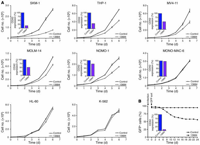
The homeobox transcription factor CDX2 plays an important role in embryonic development and regulates the proliferation and differentiation of intestinal epithelial cells in the adult. We have found that CDX2 is expressed in leukemic cells of 90% of patients with acute myeloid leukemia (AML) but not in hematopoietic stem and progenitor cells derived from normal individuals. Stable knockdown of CDX2 expression by RNA interference inhibited the proliferation of various human AML cell lines and strongly reduced their clonogenic potential in vitro. Primary murine hematopoietic progenitor cells transduced with Cdx2 acquired serial replating activity, were able to be continuously propagated in liquid culture, generated fully penetrant and transplantable AML in BM transplant recipients, and displayed dysregulated expression of Hox family members in vitro and in vivo. These results demonstrate that aberrant expression of the developmental regulatory gene CDX2 in the adult hematopoietic compartment is a frequent event in the pathogenesis of AML; suggest a role for CDX2 as part of a common effector pathway that promotes the proliferative capacity and self-renewal potential of myeloid progenitor cells; and support the hypothesis that CDX2 is responsible, in part, for the altered HOX gene expression that is observed in most cases of AML.


Figures

Figure 1
Figure 1. CDX2 expression in AML.
CDX2 mRNA levels were measured by RQ-PCR in 170 AML patients from different cytogenetic subgroups, as well as in BMMCs (n = 10), CD34+ cells (n = 3), HSCs (n = 3), CMPs (n = 3), GMPs (n = 3), and MEPs (n = 3) from normal individuals. Circles indicate patients with genomic amplification of the CDX2 locus, as assessed by aCGH and FISH (25). Bars indicate median values.
Figure 2
Figure 2. Proliferation of AML cell lines after shRNA-mediated silencing of CDX2 expression.
(A) Downregulation of CDX2 expression by shRNA TRCN13684 inhibited proliferation of the CDX2-expressing AML cell lines SKM-1, THP-1, MV4-11, MOLM-14, and NOMO-1. In MONO-MAC-6 cells, shRNA TRCN13684 did not induce efficient CDX2 mRNA knockdown, and there was no effect on cell proliferation. Similarly, treatment with shRNA TRCN13684 had no inhibitory effect in the CDX2-negative cell lines HL-60 and K-562. For each of the 6 CDX2-expressing cell lines, the degree of mRNA knockdown is shown (inset) (SKM-1, 84%; THP-1, 68%; MV4-11, 52%; MOLM-14, 49%; NOMO-1, 23%; MONO-MAC-6, 4%). Experiments were performed in triplicate. Values are represented as mean ± SEM. (B) SKM-1 cells were transduced with a lentiviral vector that coexpresses shRNA TRCN13684 and GFP. Sorted cells (proportion of GFP+ cells, 98%) were cultured at a density of 0.5 × 10⁶ to 1 × 10⁶/ml, and the GFP+ fraction was measured by flow cytometry at the indicated time points. The toxicity of CDX2 knockdown was evidenced by a relative depletion of GFP+ cells over time. In contrast, analysis of SKM-1 cells transduced with a GFP-expressing pLKO.1 construct without an shRNA sequence (proportion of GFP+ cells, 95%) showed no decrease in the percentage of GFP+ cells. The degree of CDX2 mRNA knockdown is shown (inset; 93%).
Figure 3
Figure 3. Colony formation of AML cell lines after shRNA-mediated silencing of CDX2 expression.
(A) Colony-forming assays showed a significant reduction in the number of colonies for the 5 CDX2-expressing cell lines SKM-1, MV4-11 (1 × 10⁴ plated cells), THP-1, MOLM-14, and NOMO-1 (1 × 10³ plated cells) after CDX2 mRNA knockdown by shRNA TRCN13684 as compared with cells transduced with the nonsilencing control construct. In contrast, transduction with shRNA TRCN13684 did not reduce colony formation of EOL-1, which had a very high CDX2 expression level (37,388; 82% mRNA knockdown; estimated residual expression level, 6,730); MONO-MAC-6, in which CDX2 expression was not efficiently silenced (4% mRNA knockdown); and HL-60 and K-562, which do not express CDX2 mRNA. Experiments were performed in duplicate. Values are represented as mean ± SEM. *P < 0.05; **P < 0.001. (B and C) Microscopic analysis of colonies derived from shRNA-transduced cells (right panels) showed a decrease in the number of colonies and the number of cells per colony, as compared with cells transduced with the nonsilencing control construct (left panels), for CDX2-expressing cell lines but not for CDX2-negative K-562 cells. Representative photomicrographs of methylcellulose cultures are shown. Original magnification, ×20 and ×100, respectively. (D) CDX2-expressing AML cell lines showed varying absolute mRNA levels after shRNA-mediated CDX2 knockdown.
Figure 4
Figure 4. In vitro self-renewal of murine BM and committed hematopoietic progenitor populations expressing Cdx2.
(A) Whole primary murine BM expressing Cdx2 demonstrated replating potential to the fourth plating, whereas cells transduced with empty vector had a finite ability to serially replate. Experiments were performed in duplicate. Values are represented as mean ± SD. (B) Whole BM cells derived from the fourth plating could be expanded in IL-3-supplemented liquid culture. (C) Microscopic analysis of May-Grünwald-Giemsa–stained cytospin preparations of cells derived from the fourth plating demonstrated predominantly undifferentiated myeloid morphology. Original magnification, ×1,000. (D) Flow cytometric analysis of cells derived from the fourth plating showed expression of myeloid antigens and the immaturity markers Sca-1 and c-Kit and demonstrated the absence of CD3+ or B220+ lymphoid cells and Ter119+ erythroid cells. (E) Committed murine hematopoietic progenitors and HSCs expressing Cdx2 demonstrated replating potential to the fourth plating, whereas cells transduced with empty vector had a finite ability to serially replate. Experiments were performed in duplicate. Values are represented as mean ± SD. (F) Hematopoietic progenitors and HSCs derived from the fourth plating could be expanded in IL-3–supplemented liquid culture.
Figure 6
Figure 6. Hox gene expression in murine hematopoietic cells expressing Cdx2.
(A) As compared with cells transduced with empty vector, c-Kit+Lin murine hematopoietic progenitors expressing Cdx2 demonstrated upregulation of Hoxb8 and decreased expression of Hoxa10. There were no significant changes in mRNA levels of Hoxa6, Hoxa7, and Hoxa9. No expression was detected for Hoxb3, Hoxb6, and Hoxc6 in Cdx2-transduced cells as well as in cells transduced with empty vector. For normalization, Gapdh was used. Experiments were performed in duplicate. Values are represented as mean ± SEM. (B) Spleen cells isolated from diseased secondary BM transplant recipients demonstrated upregulation of Hoxb6 and decreased expression of Hoxa7 and Hoxa9 as compared with spleen cells obtained from age-matched control mice. There were no substantial changes in mRNA levels of Hoxa6, Hoxb8, and Hoxc6. No expression was detected for Hoxa10 and Hoxb3 in spleen cells from secondary BM transplant recipients or from control mice. The expression level of Cdx2 is also indicated. For normalization, the Gapdh gene was used. Experiments were performed in duplicate. Values are represented as mean ± SEM.
Figure 5
Figure 5. Mouse model of aberrant Cdx2 expression.
(A) Mice transplanted with BM cells expressing Cdx2 (n = 5) developed AML after a median of 187 days after transplantation, whereas mice transplanted with MSCV-IRES-GFP–transduced BM (n = 3) showed no evidence of disease with a follow-up duration of more than 250 days (P = 0.013). Secondary recipients (n = 5) transplanted with BM from primary leukemic mice developed AML after a median of 52 days after transplantation. BMT, BM transplantation. (B) Diseased mice showed elevated wbc counts (primary recipients versus control mice, P = 0.09; secondary recipients versus control mice, P = 0.019) and splenomegaly (primary recipients versus control mice, P = 0.0029; secondary recipients versus control mice, P < 0.0001). Values are represented as mean ± SD. (C) Microscopic analysis of PB from primary and secondary leukemic animals demonstrated leukocytosis consisting of frequent immature myeloid cells with a high proportion of blast forms that extensively involved the BM, liver, and spleen. Panels display Wright-Giemsa–stained PB smears and H&E-stained tissue sections from representative mice transplanted with BM cells expressing Cdx2. Original magnification, ×400 and ×1,000 (PB); ×100 and ×600 (BM and liver); and ×40 and ×600 (spleen). (D) Flow cytometric analysis of GFP-gated cells from BM and spleen of primary and secondary leukemic animals demonstrated an increased proportion of Mac-1+ myeloid cells with variable expression of Gr-1, CD34, and c-Kit and a concomitant reduction in the level of CD3+ or B220+ lymphoid cells. The percentages of positive cells within the GFP+ compartment are indicated.

Comment in

References

    1. Abramovich C., Humphries R.K. Hox regulation of normal and leukemic hematopoietic stem cells. Curr. Opin. Hematol. 2005;12:210–216. - PubMed
    1. Deschamps J., et al. Initiation, establishment and maintenance of Hox gene expression patterns in the mouse. Int. J. Dev. Biol. 1999;43:635–650. - PubMed
    1. Isaacs H.V., Pownall M.E., Slack J.M. Regulation of Hox gene expression and posterior development by the Xenopus caudal homologue Xcad3. EMBO J. 1998;17:3413–3427. - PMC - PubMed
    1. van den Akker E., et al. Cdx1 and Cdx2 have overlapping functions in anteroposterior patterning and posterior axis elongation. Development. 2002;129:2181–2193. - PubMed
    1. Charite J., et al. Transducing positional information to the Hox genes: critical interaction of cdx gene products with position-sensitive regulatory elements. Development. 1998;125:4349–4358. - PubMed

Publication types