. 2007 May;205(1):154-65.
doi: 10.1016/j.expneurol.2007.01.023. Epub 2007 Feb 3.

Temporal relationship of peroxynitrite-induced oxidative damage, calpain-mediated cytoskeletal degradation and neurodegeneration after traumatic brain injury

Affiliations

Temporal relationship of peroxynitrite-induced oxidative damage, calpain-mediated cytoskeletal degradation and neurodegeneration after traumatic brain injury

Ying Deng et al. Exp Neurol. 2007 May.

Abstract

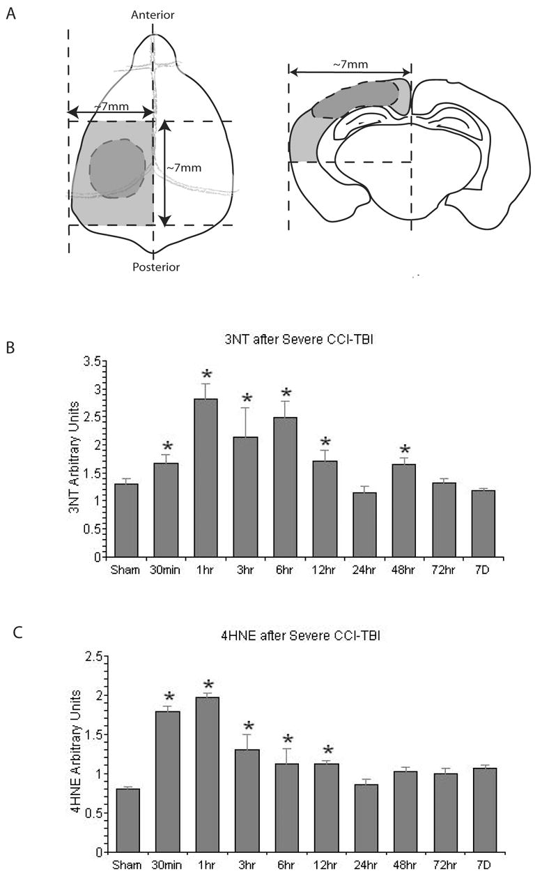
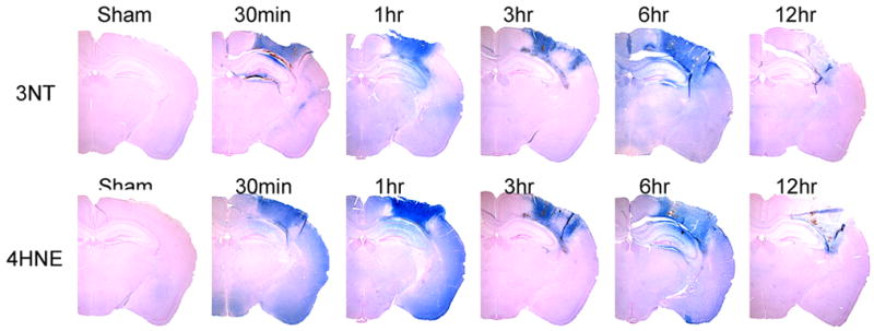
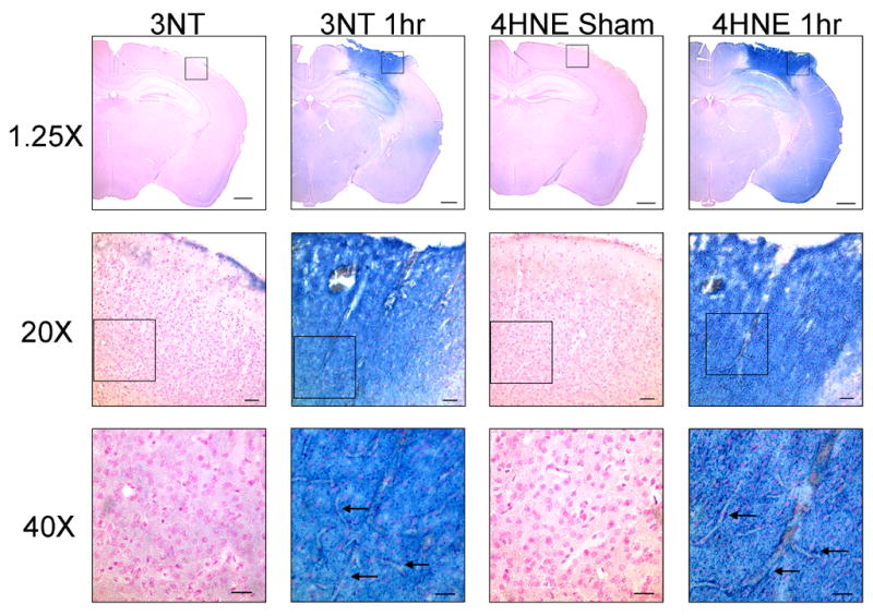
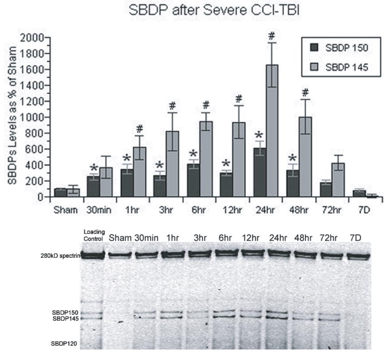
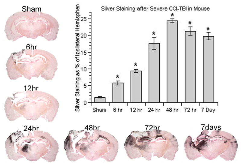
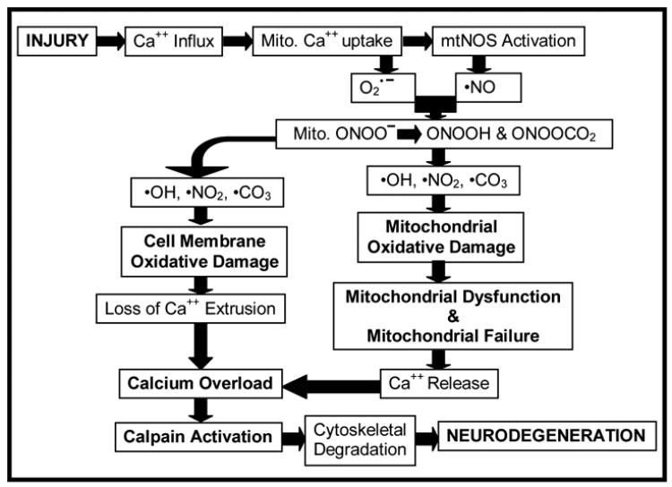
We assessed the temporal and spatial characteristics of PN-induced oxidative damage and its relationship to calpain-mediated cytoskeletal degradation and neurodegeneration in a severe unilateral controlled cortical impact (CCI) traumatic brain injury (TBI) model. Quantitative temporal time course studies were performed to measure two oxidative damage markers: 3-nitrotyrosine (3NT) and 4-hydroxynonenal (4HNE) at 30 min, 1, 3, 6, 12, 24, 48, 72 h and 7 days after injury in ipsilateral cortex of young adult male CF-1 mice. Secondly, the time course of Ca(++)-activated, calpain-mediated proteolysis was also analyzed using quantitative western-blot measurement of breakdown products of the cytoskeletal protein alpha-spectrin. Finally, the time course of neurodegeneration was examined using de Olmos silver staining. Both oxidative damage markers increased in cortical tissue immediately after injury (30 min) and elevated for the first 3-6 h before returning to baseline. In the immunostaining study, the PN-selective marker, 3NT, and the lipid peroxidation marker, 4HNE, were intense and overlapping in the injured cortical tissue. alpha-Spectrin breakdown products, which were used as biomarker for calpain-mediated cytoskeletal degradation, were also increased after injury, but the time course lagged behind the peak of oxidative damage and did not reach its maximum until 24 h post-injury. In turn, cytoskeletal degradation preceded the peak of neurodegeneration which occurred at 48 h post-injury. These studies have led us to the hypothesis that PN-mediated oxidative damage is an early event that contributes to a compromise of Ca(++) homeostatic mechanisms which causes a massive Ca(++) overload and calpain activation which is a final common pathway that results in post-traumatic neurodegeneration.


Figures

Figure 1
Figure 1
Slot-blotting studies in ipsilateral cortical traumatic brain injury tissues showing the temporal changes in protein nitration (3-nitrotyrosine; 3NT) and lipid peroxidation. A. Schematic showing peri-contusional cortical tissue samples; B. time course of changes in 3-NT; C. Time course of LP end product 4HNE. N = 8 animals per timepoint; values = mean ± standard error; one-way ANOVA and Fisher’s PLSD post hoc test: *P < 0.0001 vs. Sham.
Figure 2
Figure 2
Immunohistochemical (IHC) staining studies showing the time course and spatial extent of PN-induced 3NT and 4HNE at the epicenter (Bregma-2.0mm) of the injury site during the first 12 hrs compared to Sham. The IHC staining indicated prominent increase of both markers after injury and similar distribution between the two.
Figure 3
Figure 3
High power photomicrographs of 3NT and 4HNE immunostaining in the contusion site in a sham, non-injured brain compared to staining at the contusion site at 1 hr after injury. At 20x and 40x magnification, intense staining for both oxidative damage markers is seen throughout the neuropil. In addition, microvascular staining for both 3NT and 4HNE is seen to clearly outline the microvessels (arrows) deep within the injured cortex. Although the Sham brain shows some light staining in the neuropil, there is no evidence of staining of non-injured microvessels. Please note that the 20x and 40x focus was adjusted to emphasize the microvascular staining, and as a result the background tissue appears slightly out of focus. The calibration bar for the 1.25x photomicrographs is 2.0mm; for 20x the calibration bar is 100μm; for 40x the calibration bar is 50μm.
Figure 4
Figure 4
Time course of the post-traumatic increase in calpain-mediated α-spectrin breakdown products in CCI cortical tissues. Spectrin breakdown product (SBDP) 145 is specific to calpain activity, whereas SBDP 150 is produced by both calpain and caspase 3. Both SBDPs showed similar patterns, except that calpain-mediated SBDP 145 showed a more prominent increase. Both SBDPs have an immediate increase following injury, and did not reach their peak until 24 hrs. N = 8 animals per timepoint; values = mean + standard error; one-way ANOVA and Fisher’s PLSD post hoc test: *P < 0.0001 vs. Sham SBDP150; one-way ANOVA and Fisher’s PLSD post hoc test: #P < 0.0001 vs. Sham SBDP145.
Figure 5
Figure 5
Time course of post-traumatic neurodegeneration in CCI model as revealed by the de Olmos silver staining technique. The bar chart provides the quantification of the lesion volume abut measurement of the silver staining. An increase in silver staining volume determined by image analysis (see Materials and Methods) occurred as early as 6 hrs and reached its peak at 48 hrs post-injury. N = 3–4 animals per time point; values = mean ± standard error; one-way ANOVA and Fisher’s PLSD post hoc test: *P < 0.0001 vs. Sham.
Figure 6
Figure 6
Hypothetical interrelationship between PN-induced oxidative damage in neuronal mitochondria and the rest of the neuron, compromise of Ca++ homeostasis, calpain-mediated proteolysis and neurodegeneration. Our results suggest that PN-induced oxidative damage plays a key role in the post-traumatic secondary injury, which leads to exacerbation of Ca++ overload, calpain proteolysis of cytoskeletal and other cellular proteins and neurodegeneration. See Discussion for a further description.

References

    1. Arrigoni E, Cohadon F. Calcium-activated neutral protease activities in brain trauma. Neurochem Res. 1991;16:483–487. - PubMed
    1. Awasthi D, Church DF, Torbati D, Carey ME, Pryor WA. Oxidative stress following traumatic brain injury in rats. Surg Neurol. 1997;47:575–581. discussion 581–572. - PubMed
    1. Bartus RT, Elliott PJ, Hayward NJ, Dean RL, Harbeson S, Straub JA, Li Z, Powers JC. Calpain as a novel target for treating acute neurodegenerative disorders. Neurol Res. 1995;17:249–258. - PubMed
    1. Bates TE, Loesch A, Burnstock G, Clark JB. Immunocytochemical evidence for a mitochondrially located nitric oxide synthase in brain and liver. Biochem Biophys Res Commun. 1995;213:896–900. - PubMed
    1. Beckman JS. Oxidative damage and tyrosine nitration from peroxynitrite. Chem Res Toxicol. 1996;9:836–844. - PubMed

MeSH terms