. 2007 Mar 9;128(5):947-59.
doi: 10.1016/j.cell.2007.01.042.

An Nkx2-5/Bmp2/Smad1 negative feedback loop controls heart progenitor specification and proliferation

Affiliations

An Nkx2-5/Bmp2/Smad1 negative feedback loop controls heart progenitor specification and proliferation

Owen W J Prall et al. Cell. .

Abstract

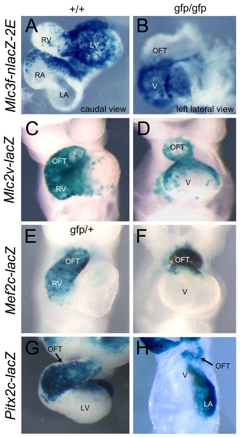
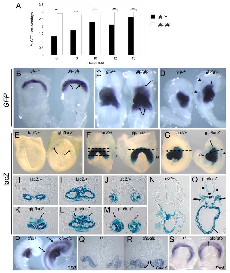
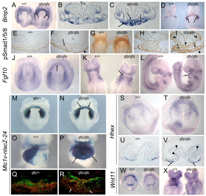
During heart development the second heart field (SHF) provides progenitor cells for most cardiomyocytes and expresses the homeodomain factor Nkx2-5. We now show that feedback repression of Bmp2/Smad1 signaling by Nkx2-5 critically regulates SHF proliferation and outflow tract (OFT) morphology. In the cardiac fields of Nkx2-5 mutants, genes controlling cardiac specification (including Bmp2) and maintenance of the progenitor state were upregulated, leading initially to progenitor overspecification, but subsequently to failed SHF proliferation and OFT truncation. In Smad1 mutants, SHF proliferation and deployment to the OFT were increased, while Smad1 deletion in Nkx2-5 mutants rescued SHF proliferation and OFT development. In Nkx2-5 hypomorphic mice, which recapitulate human congenital heart disease (CHD), OFT anomalies were also rescued by Smad1 deletion. Our findings demonstrate that Nkx2-5 orchestrates the transition between periods of cardiac induction, progenitor proliferation, and OFT morphogenesis via a Smad1-dependent negative feedback loop, which may be a frequent molecular target in CHD.


Figures

Figure 1
Figure 1
Analysis of FHF and SHF contributions to Nkx2-5 null embryos. A–H. Expression of indicated lacZ transgenes in wildtype (+/+) and Nkx2-5gfp/gfp null (gfp/gfp) embryos at E9.0. Abbreviations: OFT: outflow tract; LV: left ventricle; LA: left atrium; RA: right atrium; RV: right ventricle; V: ventricle-like chamber in null embryos.
Figure 2
Figure 2
Microarray analysis. A. Fluorescence channels 1 (Fl1, 530 nM) and 2 (Fl2, 585 nM) FACS plots of wildtype (+/+), Nkx2-5gfp/+ heterozygous and Nkx2-5gfp/gfp null embryos (both axes log10 scale). Gates define propidium iodide-permeable dead cells (dashed line) and live Nkx2-5-GFP+ cells (solid line). Nkx2-5-GFP+ cells (green) were used for microarray analysis. B. Microarray heat map showing fold-change in mRNA expression between Nkx2-5 null and heterozygous GFP+ cells (see scale). Genes modified at least 1.7-fold at 3 embryonic stages were clustered by standard correlation analysis (see tree diagrams for genes, left, and for embryonic stages, above). Genes known to be down-regulated in Nkx2-5 mutants are arrowed. The prominent cluster of genes up-regulated from 6–21 ps is indicated (*).
Figure 3
Figure 3
Cardiac progenitor genes/proteins up-regulated in Nkx2-5 null embryos. A–X. Whole mount in situ hybridization and immunohistochemistry with representative sections showing expression of indicated cardiac progenitor cell genes/proteins in wildtype (+/+) and Nkx2-5gfp/gfp embryos at E7.5–E8.5. Regions of up-regulated expression in Nkx2-5 mutants are indicated (arrows, cardiac mesoderm; arrowheads, endoderm).
Figure 4
Figure 4
Over-specification of cardiac progenitors in Nkx2-5 null embryos. A. Percentage of Nkx2-5-GFP+ cells/embryo in Nkx2-5gfp/+ control and Nkx2-5gfp/gfp embryos detected by FACS (6–15ps). An average of ~7,200 cells (6–10ps) and 23,000 cells (12–15ps) were counted for each sample. Asterisks show significance by Chi squared analysis (*** p<0.001, ** p<0.01, * p<0.05). B–D. GFP mRNA expression in Nkx2-5gfp/+ and Nkx2-5gfp/gfp null embryos at E7.75 (B) and E8.5 (C,D). Ectopic Nkx2-5-GFP expression is indicated in tissues adjacent to the heart (arrows) and in neural tube (arrowheads). E–O. Xgal staining for Nkx2-5-lacZ expression in Nkx2-5lacZ/+ control and Nkx2-5gfp/lacZ null embryos at E7.5 (E), E8.0 (F and H–M) and E9.5 (G,N,O). Ectopic Nkx2-5-lacZ expression in the cardiac region (arrows), pharyngeal mesenchyme (double arrows), neural tube (arrowheads) and foregut (double arrowheads) is indicated. Dashed lines in F and G indicate planes of sections in H–M and N,O, respectively. P. GFP mRNA expression at E9.0 in Nkx2-5gfp/+ and Nkx2-5gfp/gfp embryos showing induction in neural tube and head mesoderm remote from the cardiac region in mutants. Q,R Expanded expression of Gata4 at E8.0 in the medial wall of the intraembryonic coelom (arrows) in Nkx2-5gfp/gfp embryos. S. Expanded expression of Tbx5 at E7.75 in Nkx2-5gfp/gfp embryos (arrow).
Figure 5
Figure 5
Modified expression of cardiac signaling components in Nkx2-5 null embryos. AD. Bmp2 expression in wildtype (+/+) and Nkx2-5gfp/gfp null embryos at E8.0 (A–C) and E8.5 (D). Elevated expression in mutants arrowed. E–I. Phospho-Smad1/5/8 expression at E7.75 (E,F) and E8.0 (G–I). Elevated expression in mutants indicated in cardiac mesoderm (single arrows), endoderm (arrowheads) and ventral neural tube (double arrows). J–L. Fgf10 expression at E7.5 (J), E8.5 (K) and E9.5 (L), arrows showing ectopic expression. M–P.β-galactosidase activity from the Mlc1v-nlacZ-24 transgene at E8.0 (M,N) and E9.5 (O,P), with arrows showing ectopic expression. Q,R. Co-immuno-detection of β-galactosidase protein from the Mlc1v-nlacZ-24 transgene (green nuclei) and sarcomeric myosin (red) at E7.5 in the cardiac crescent in Nkx2-5gfp/+ control and Nkx2-5gfp/gfp embryos. Arrows indicate expanded expression of β-galactosidase in myosin+ cardiomyocytes in mutants. S–V. Hhex expression at E7.75 showing elevated expression in cardiac endoderm (arrows) and endothelial cells within cranial mesenchyme (arrowheads) of Nkx2-5gfp/gfp embryos. W,X. Wnt11 expression (arrows) is severely reduced in Nkx-2-5 null embryos at E7.75 (Y) and E8.5 (Z).
Figure 6
Figure 6
Nkx2-5 and Smad1 have opposing effects on SHF proliferation and deployment. A,B. Cell proliferation in the SHF of Nkx2-5gfp/gfp mutant versus wildtype control (+/+) embryos (A, E8.0, 6–8ps, C57BL/6 genetic background) and embryos deleted for Smad1 in mesoderm (Mesp1Cre/+/Smad1fl/fl) versus heterozygous deleted controls (Mesp1Cre/+/Smad1fl/+) (B, E8.0, 6–8ps, 75% Quackenbush-Swiss/25% C57BL/6) and for littermate embryos from Nkx2-5+/gfp X Smad1+/ crosses (C, E8.5, 10–12ps, 50% Quackenbush-Swiss/50% C57BL/6). Shown is mean % of phosphohistone H3+ (pHH3) cells +/− standard deviation, n=3 for each genotype. D–F. ISH for Wnt11 mRNA highlighting increased OFT/RV (arrows) size in Mesp1Cre/+/Smad1fl/fl embryos versus Mesp1+/+/Smad1fl/fl and Mesp1Cre/+/Smad1fl/+ controls at E9.5. G–V, Marker analysis of wildtype, Nkx2-5 null, Smad1 null and Nkx2-5/Smad1 double null littermate embryos for Bmp2 (E8.0; G–J), Nkx2-5-GFP (E8.25; sections, K–N), Wnt11 (E8.5; ventral views, O–R; right-hand side views, O′–R′; section through OFT, O″–R″) and Isl1 (E8.25; sections, S–V. Note in Fig. 6L the expanded Nkx2-5-GFP expression in the dorsal pericardium (double arrows), adjacent mesenchyme (arrow), dorsal foregut (arrowhead) and ventral neural tube (double arrowheads) of Nkx2-5/Smad1 double null embryos. In Fig. 6O–R″ Wnt11 expression is indicated in the OFT (arrows) and inflow region (arrowheads) of wildtype (O,O′,O″), Smad1 null (Q,Q′,Q″) and Nkx2-5/Smad1 double null (R,R′,R″) embryos but is absent from the rudimentary OFT of Nkx2-5 null embryos (outlined by dotted lines, P,P′,P″). Persistent Isl1 expression in cardiomyocytes located in the heart tubes of Nkx2-5 null (T) and Nkx2-5/Smad1 double null (V) embryos is indicated (arrows).
Figure 7
Figure 7
Genetic rescue of congenital cardiac malformations in Nkx2-5 hypomorphic embryos. A–D. Gross morphology of Nkx2-5 hypomorphic embryos (gfp/IRES-cre) and littermate controls (+/IRES-cre) at E17.5 showing malrotation and rightward displacement of the aorta (dashed lines) and pulmonary artery (solid lines) in hypomorphs (A), with sections from E14.5 embryos showing DORV (C) or OA with large VSD (D). E–H. Morphological analysis of early OFT development showing compromised outflow development (arrows) in hypomorphs. Nkx2-5 expression highlights cardiac structures at E8.5 (E,F). Wnt11 expression marks the RV/OFT at E9.5 (G,H). I–K. Molecular analysis of cardiac progenitor cell defects in Nkx2-5gfp/IRES-cre hypomorphic embryos and Nkx2-5+/IRES-cre controls. Note up-regulated expression of cardiac progenitor cell markers Bmp2 (I, E7.75) and Isl1 (J,K, E7.5) in hypomorphs (arrows, pre-cardiac mesoderm; arrowheads, endoderm). L, Decreased proliferation in the SHF of Nkx2-5gfp/IRES-cre hypomorphic embryos (E8.5, 10–12ps, C57BL/6 genetic background, mean +/− SD, n=3 for each genotype). M–X, Rescue of early OFT defects in Nkx2-5gfp/IRES-cre hypomorphic embryos by deletion of a single allele of Smad1. The immature AV cushions in hypomorphs are indicated by an asterisk (S,T). Abbreviations: Ao: aortic root; ASD: atrial septal defect; DORV: double outlet right ventricle; LV: left ventricle; MV: mitral valve; OA: overriding aorta; PA: pulmonary artery; RV: right ventricle; TV: tricuspid valve; VSD: ventricular septal defect.

References

    1. Benson DW, Silberbach GM, Kavanaugh-McHugh A, Cottrill C, Zhang Y, Riggs S, Smalls O, Johnson MC, Watson MS, Seidman JG, et al. Mutations in Nkx2-5, a Cardiac Transcription Factor, Affect Diverse Cardiac Developmental Pathways. J Clin Invest. 1999;104:1567–1573. - PMC - PubMed
    1. Biben C, Harvey RP. Homeodomain factor Nkx2-5 controls left-right asymmetric expression of bHLH eHand during murine heart development. Genes Dev. 1997;11:1357–1369. - PubMed
    1. Biben C, Weber R, Kesteven S, Stanley E, McDonald L, Elliott DA, Barnett L, Koentgen F, Robb L, Feneley M, Harvey RP. Cardiac septal and valvular dysmorphogenesis in mice heterozygous for mutations in the homeobox gene Nkx2-5. Circ Res. 2000;87:888–895. - PubMed
    1. Buckingham M, Meilhac S, Zaffran S. Building the mammalian heart from two sources of myocardial cells. Nat Rev Genet. 2005;6:826–835. - PubMed
    1. Cai CL, Liang X, Shi Y, Chu PH, Pfaff SL, Chen J, Evans S. Isl1 identifies a cardiac progenitor population that proliferates prior to differentiation and contributes a majority of cells to the heart. Dev Cell. 2003;5:877–889. - PMC - PubMed

Publication types

MeSH terms