Comparative Study
. 2007 May 1;305(1):120-32.
doi: 10.1016/j.ydbio.2007.02.002. Epub 2007 Feb 7.

Sox9 is required for precursor cell expansion and extracellular matrix organization during mouse heart valve development

Affiliations
Comparative Study

Sox9 is required for precursor cell expansion and extracellular matrix organization during mouse heart valve development

Joy Lincoln et al. Dev Biol. .

Abstract

Heart valve structures derived from mesenchymal cells of the endocardial cushions (ECs) are composed of highly organized cell lineages and extracellular matrix. Sox9 is a transcription factor required for both early and late stages of cartilage formation that is also expressed in the developing valves of the heart. The requirements for Sox9 function during valvulogenesis and adult valve homeostasis in mice were examined by conditional inactivation of Sox9 using Tie2-cre and Col2a1-cre transgenes. Sox9(flox/flox);Tie2-cre mice die before E14.5 with hypoplastic ECs, reduced cell proliferation and altered extracellular matrix protein (ECM) deposition. Sox9(flox/flox);Col2a1-cre mice die at birth with thickened heart valve leaflets, reduced expression of cartilage-associated proteins and abnormal ECM patterning. Thickened valve leaflets and calcium deposits, characteristic of valve disease, are observed in heterozygous adult Sox9(flox/+);Col2a1-cre mice. Therefore, Sox9 is required early in valve development for expansion of the precursor cell population and later is required for normal expression and distribution of valvular ECM proteins. These data indicate that Sox9 is required for early and late stages of valvulogenesis and identify a potential role for Sox9 in valve disease mechanisms.


Figures

Figure 1
Figure 1. Sox9 and Cartilage Link Protein are expressed in endocardial cushions and primitive heart valve leaflet structures during mouse valvulogenesis
Immunohistochemistry was used to examine expression patterns of Sox9 (A, C) and CLP (B, D) in the endocardial cushions at E13.5 (A, B) and developing heart valve leaflets at E18.5 (C, D). (A) Nuclear Sox9 expression is detected throughout valve precursor cells of the central (arrows) and mural (arrowhead) ECs. (B) CLP expression is also detected in the matrix associated with Sox-9 positive cells within the mural EC (arrowhead) and the more lateral ventricular region of the central EC (arrows). At E18.5, Sox9 (C) and CLP (D) expression are detected in the maturing septal and mural tricuspid valve leaflets (arrows), with predominant expression observed on the atrial surface (arrow) and ventricular surface with enhanced expression at the distal tips (arrowheads). Notably, Sox9 and CLP expression is not detected in the myocardium (*). tv, tricuspid valve; IVS, interventricular septum; RA, right atrium; LV, left ventricle; CLP, Cartilage Link Protein.
Figure 2
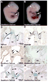
Figure 2. Sox9flox/flox;Tie2-cre mice have embryonic heart failure and display hypoplastic endocardial cushions
(B) Grossly, Sox9flox/flox;Tie2-cre mice display pericardial edema (arrow) and increased blood pooling (arrowhead), compared to controls (A) at E12.5. At the cellular level, X-gal staining and in situ hybridization were performed on tissue sections of heart valve structures at E13.5 from Tie2-cre mice bred with ROSA26R to determine Tie2-cre-mediated recombination with Sox9 in the developing heart valves. (C) Tie2-cre recombination is detected throughout the valve primordia at E13.5, consistent with Sox9 expression (arrows, D). (E) Compared to control Sox9flox/+;Tie2-cre control mice, Sox9 protein expression (arrows) is diminished in ECs of Sox9flox/flox;Tie2-cre mice (F) indicative of recombination. Alcian blue staining was used to indicate proteoglycans within the heart valve structures in control Sox9flox/+;Tie2-cre (G) and mutant Sox9flox/flox;Tie2-cre (H) mice. Arrows indicate hypoplastic EC in Sox9flox/flox;Tie2-cre mice (D) compared to controls (C). Arrowheads indicate incomplete formation of the atrial septum (as). IVS, interventricular septum; mv, mitral valve; tv, tricuspid valve; LV, left ventricle; RV, right ventricle.
Figure 3
Figure 3. Heart valve leaflets in Sox9flox/flox;Col2a1-cre mice are abnormal with reduced Alcian blue staining
Col2a1-cre recombination (A) is observed in a subset of cells on the atrial (A) and ventricular (V) surfaces of the valve leaflets at E18.5 in Col2a1-cre mice bred with Rosa26R. (B) At this time, Sox9 expression is detected by in situ hybridization throughout the valve leaflet structures (arrows). (C) Compared to Sox9flox/+;Col2a1-cre control mice, nuclear Sox9 protein expression is diminished on the ventricular aspect of valve leaflets of Sox9flox/flox;Col2a1-cre mice (arrow) (D), although expression remains on the atrial surface where recombination has not taken place (arrowhead). Heart valve leaflets of Sox9flox/flox;Col2a1-cre mice (F) are abnormal at E18.5 and Alcian blue staining is reduced compared to Sox9flox/+;Col2a1-cre control mice (E). IVS, interventricular septum; mv, mitral valve; tv, tricuspid valve.
Figure 4
Figure 4. Valve precursor cells of Sox9flox/flox;Tie2-cre mice have decreased proliferation and perturbed cell lineage diversification
Alcian blue staining of histological sections of E11.5 Sox9flox/+;Tie2-cre (A) and Sox9flox/flox;Tie2-cre (B) embryos shows abnormal morphology and reduced size of EC lacking Sox9 expression indicated by arrows. Phospho-histone H3 (pHH3) immunohistochemistry was used to detect proliferating cells in EC tissue sections from Sox9flox/+;Tie2-cre (D) and Sox9flox/flox;Tie2-cre (E) mice at E12.5. (C) Quantitative analyses were performed to detect significant changes in the average number of mesenchymal cells in tissue sections of the EC from Sox9flox/flox;Tie2-cre mice compared to Sox9flox/+;Tie2-cre controls at E11.5 and E12.5. (F) The number of nuclei stained positive for pHH3 in each tissue section of EC from E12.5 Sox9flox/+;Tie2-cre and Sox9flox/flox;Tie2-cre mice was used to calculate a proliferative index represented as a percentage of the total number of nuclei. Note proliferation is reduced in Sox9flox/flox;Tie2-cre mice. (F). A, atrium; V, ventricle; RA, right atrium; IVS, interventricular septum.
Figure 5
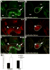
Figure 5. Tenascin and CLP expression is altered in Sox9flox/flox;Tie2-cre mice
(A, B) Immunofluorescence staining of tenascin (A, B) and CLP (C, D) were used to show ECM protein expression in tissue sections of ECs (highlighted by white lines) from Sox9flox/+;Tie2-cre (A, C, E) and Sox9flox/flox;Tie2-cre (B, D, F) mice at E12.5. Tenascin-positive regions are indicated by arrows and CLP-positive regions are indicated by arrowheads. (D) The atrial aspect of the central EC where CLP is not expressed is indicated by (*). Merged images from (A-D) are shown in (E, F). The percent area of the immunoreactivity of CLP and tenascin within the central EC is reported in (G). Error bars represent standard error of the mean, and statistical significance (*) is calculated by Student’s t-test compared to Sox9flox/+;Tie2-cre mice (P<0.05; n=3). IVS, interventricular septum; LA, left atrium; RV, right ventricle.
Figure 6
Figure 6. Type II collagen and CLP expression are reduced in Sox9flox/+;Col2a1-cre mice
Immunostaining for type II collagen (A, B), CLP (C, D) and elastin (E, F) was used to determine ECM expression and organization in Sox9flox/+;Col2a1-cre (A, C, E) and Sox9flox/flox;Col2a1-cre (B, D, F) mitral valve mural and septal leaflets at E18.5 (outlined in white). The expression of type II collagen (A) and CLP (C) in the mural and septal valve leaflets (arrows) of Sox9flox/+;Col2a1-cre mice is diminished in Sox9flox/flox;Col2a1-cre mice (B, D). (E) Normal elastin expression is detected on the atrial surface of mitral valve mural and septal leaflets of Sox9flox/+;Col2a1-cre embryos. (F) Elastin expression is also observed on the atrial surface of valve leaflets from Sox9flox/flox;Col2a1-cre embryos, but ectopic expression is additionally noted on the ventricular surface of the septal leaflet (arrowhead, F). mv, mitral valve; IVS, interventricular septum.
Figure 7
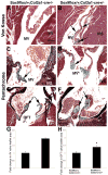
Figure 7. Heart valves from adult Sox9flox/+;Col2a1-cre mice are thickened and calcified
Von Kossa stain was used to examine calcium deposition in mitral valve (MV) leaflets of control Sox9flox/+;Col2a1-cre−/− (A) and Sox9flox/+;Col2a1-cre+/− (B) mice at 4 months of age. Von Kossa stains calcium deposits black, as indicated in Sox9flox/+;Col2a1-cre+/− mice (arrows, B). Movat’s Pentachrome staining was used to show ECM distribution and organization in the MV (C, D) and semilunar (OFT) (E, F) valve leaflets of control and Sox9flox/+;Col2a1-cre+/− mice. Morphometric analyses show thickened valve leaflets in Sox9flox/+;Col2a1-cre+/− (D, F) mice compared to controls (C, E). Quantitative analysis of the fold change in the AV (G) and OFT (H) leaflet area (outlined in black) of Sox9flox/+;Col2a1-cre mice over controls is shown. Statistical significance (*) was performed by Student’s t-test (P < 0.01). MV, mitral valve; IVS, interventricular spetum; OFT, outflow tract.

Similar articles

Cited by

References

    1. Akiyama H, Chaboissier M-C, Behringer RR, Rowitch DH, Schedl A, Epstein JA, de Crombrugghe B. Essential role of Sox9 in the pathway that controls formation of cardiac valves and septa. Proc Nat Acad Sci. 2004;101:6502–6507. - PMC - PubMed
    1. Akiyama H, Chaboissier MC, Martin JF, Schedl A, de Crombrugghe B. The transcription factor Sox9 has essential roles in successive steps of the chondrocyte differentiation pathway and is required for expression of Sox5 and Sox6. Genes Dev. 2002;16:2813–28. - PMC - PubMed
    1. Armstrong EJ, Bischoff J. Heart valve development: Endothelial cell signaling and differentiation. Circ Res. 2004;95:459–470. - PMC - PubMed
    1. Baudino T, Carver W, Giles WR, Borg TK. Cardiac Fibroblasts: friend or foe? Am J Physiol Heart Circ Physiol. 2006;291:H1015–26. - PubMed
    1. Bell DM, Leung KK, Wheatley SC, Ng LJ, Zhou S, Ling KW, Sham MH, Koopman P, Tam PP, Cheah KS. SOX9 directly regulates the type-II collagen gene. Nat Genet. 1997;16:174–8. - PubMed

Publication types

MeSH terms

Substances