. 2007 May 15;92(10):3674-85.
doi: 10.1529/biophysj.106.101238. Epub 2007 Mar 9.

Unfolding of beta-sheet proteins in SDS

Affiliations

Unfolding of beta-sheet proteins in SDS

Mette M Nielsen et al. Biophys J. .

Abstract

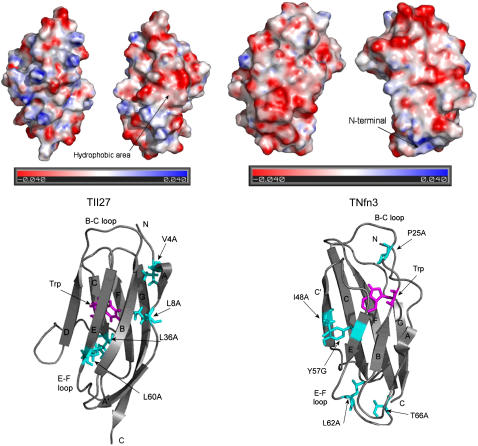
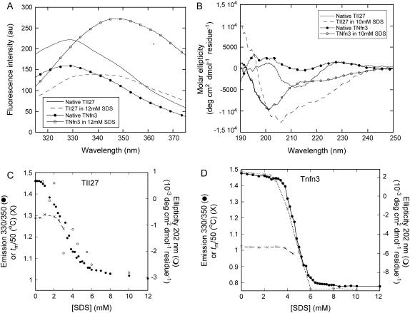
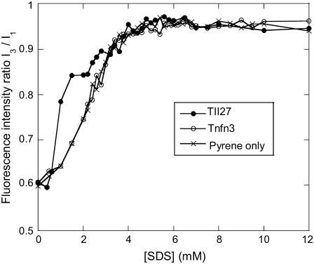
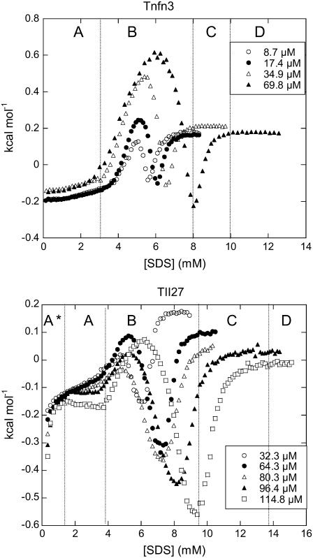
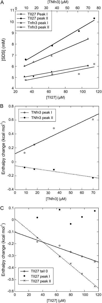
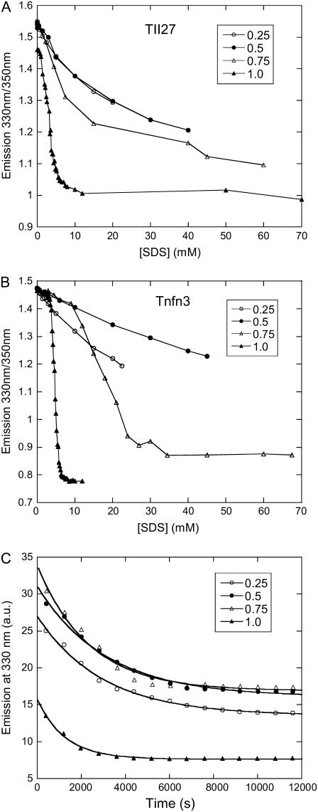
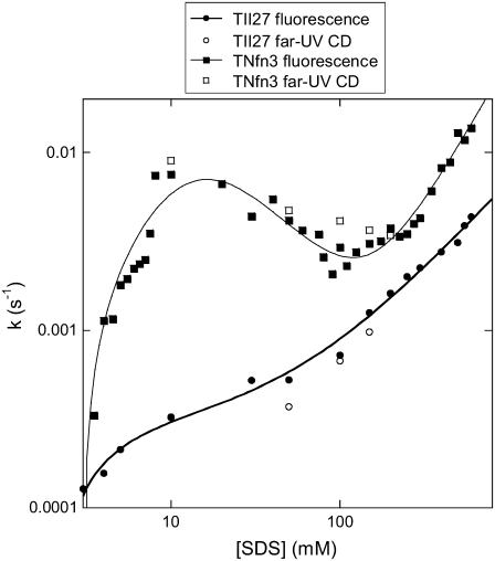
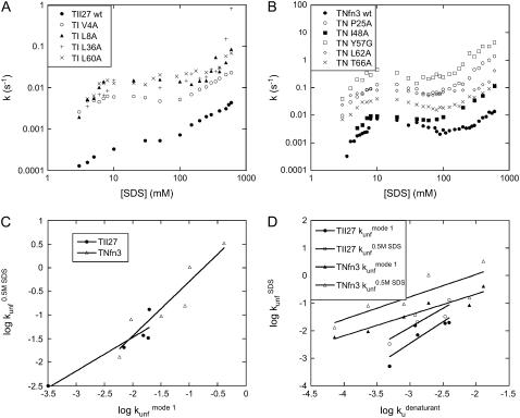
Beta-sheet proteins are particularly resistant to denaturation by sodium dodecyl sulfate (SDS). Here we compare unfolding of two beta-sandwich proteins TNfn3 and TII27 in SDS. The two proteins show different surface electrostatic potential. Correspondingly, TII27 unfolds below the critical micelle concentration via the formation of hemimicelles on the protein surface, whereas TNfn3 only unfolds around the critical micelle concentration. Isothermal titration calorimetry confirms that unfolding of TII27 sets in at lower SDS concentrations, although the total number of bound SDS molecules is similar at the end of unfolding. In mixed micelles with the nonionic detergent dodecyl maltoside, where the concentration of monomeric SDS is insignificant, the behavior of the two proteins converges. TII27 unfolds more slowly than TNfn3 in SDS and follows a two-mode behavior. Additionally TNfn3 shows inhibition of SDS unfolding at intermediate SDS concentrations. Mutagenic analysis suggests that the overall unfolding mechanism is similar to that observed in denaturant for both proteins. Our data confirm the kinetic robustness of beta-sheet proteins toward SDS. We suggest this is related to the inability of SDS to induce significant amounts of alpha-helix structure in these proteins as part of the denaturation process, forcing the protein to denature by global rather than local unfolding.


Figures

FIGURE 1
FIGURE 1
(A) Electrostatic potential of TII27 and TNfn3, illustrated using PyMOL. (B) Structures of TII27 and TNfn3, highlighting the side chains mutated in this study.
FIGURE 2
FIGURE 2
(A) Fluorescence and (B) Far-UV CD spectra of TII27 and TNfn3 in the absence and presence of SDS. (C) Equilibrium denaturation of TII27 and (D) TNfn3 followed by fluorescence, CD, and changes in the melting temperature as a function of SDS concentration. For both proteins, tm has been divided by 50 to be compatible with the fluorescence axis.
FIGURE 3
FIGURE 3
Pyrene fluorescence as a function of SDS in the absence and presence of TII27 and TNfn3. Note the steep increase in the I3/I1 ratio of TII27 between 0.5 and 3 mM SDS compared to protein-free pyrene and pyrene in the presence of TNfn3. This suggests that hemimicellar structures form on the TII27 surface but not on TNfn3.
FIGURE 4
FIGURE 4
ITC enthalpograms of (left) Tnfn3 and (right) TII27 upon titration with SDS. See text for an explanations of sections A*–D.
FIGURE 5
FIGURE 5
(A) Concentrations at which peaks I and II appear for TII27 and TNfn3 as a function of protein concentration. (B and C) Enthalpy values at peaks I and II as well as in region A* (for TII27) as a function of protein concentration. Data are summarized in Table 1.
FIGURE 6
FIGURE 6
Unfolding of (A) TII27 and (B) TNfn3 in mixed micelles of SDS and DDM at SDS mole fractions 0.25–1 followed by the change in emission ratio 330:350 nm. (C) Kinetics of unfolding of TII27 in 80 mM detergent containing 0.25–1 mole fraction SDS.
FIGURE 7
FIGURE 7
Rate constants of unfolding of TII27 and TNfn3 as a function of SDS concentration. Data for TII27 are fitted to mode 1 + 3 unfolding, whereas data for TNfn3 are fitted to mode 1 + 2 + 3 unfolding (Scheme 3).
FIGURE 8
FIGURE 8
Unfolding rate constants for mutants of (A) TII27 and (B) TNfn3 as a function of SDS. Data are summarized in Table 2. Correlation of unfolding rate constants in SDS (C) with each other and (D) with unfolding rate constants in buffer extrapolated from unfolding rate constants in denaturant. The slopes, intercepts, and correlation coefficients for the best linear fits are summarized in Table 3.

References

    1. La Mesa, C. 2005. Polymer-surfactant and protein-surfactant interactions. J. Coll. Int. Sci. 286:148–157. - PubMed
    1. Timasheff, S. 2002. Protein hydration, thermodynamic binding, and preferential hydration. Biochemistry. 41:13473–13482. - PubMed
    1. Kirk, O., T. V. Borchert, and C. C. Fuglsang. 2002. Industrial enzyme applications. Curr. Opin. Biotechnol. 13:345–351. - PubMed
    1. Sehgal, P., and D. E. Otzen. 2006. Thermodynamics of unfolding of an integral membrane protein in mixed micelles. Prot. Sci. 15:890–899. - PMC - PubMed
    1. Otzen, D. E., L. Christiansen, and M. Schülein. 1999. A comparative study of the unfolding of the endoglucanase Cel45 from Humicola insolens in denaturant and surfactant. Prot. Sci. 8:1878–1887. - PMC - PubMed

Publication types

LinkOut - more resources