. 2007 Mar;19(3):890-903.
doi: 10.1105/tpc.106.048058. Epub 2007 Mar 9.

Impairment of cellulose synthases required for Arabidopsis secondary cell wall formation enhances disease resistance

Affiliations

Impairment of cellulose synthases required for Arabidopsis secondary cell wall formation enhances disease resistance

Camilo Hernández-Blanco et al. Plant Cell. 2007 Mar.

Abstract

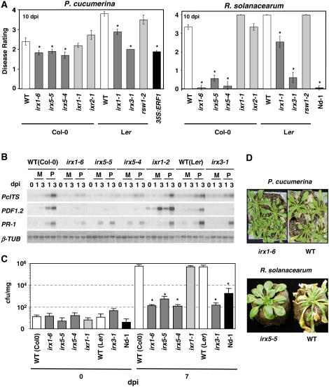
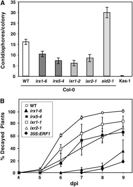
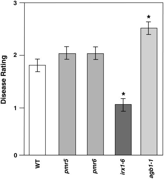
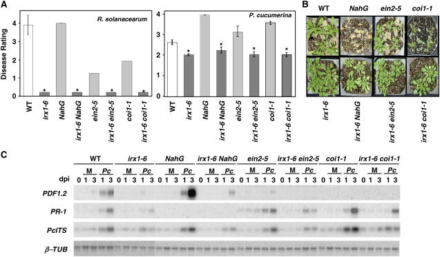
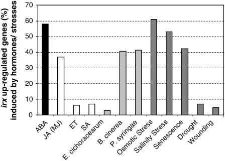
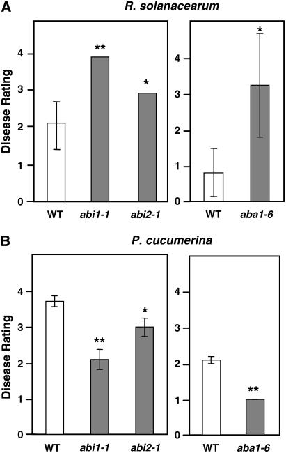
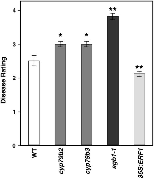
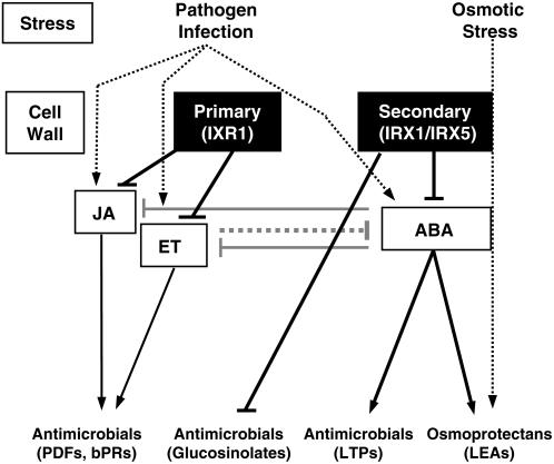
Cellulose is synthesized by cellulose synthases (CESAs) contained in plasma membrane-localized complexes. In Arabidopsis thaliana, three types of CESA subunits (CESA4/IRREGULAR XYLEM5 [IRX5], CESA7/IRX3, and CESA8/IRX1) are required for secondary cell wall formation. We report that mutations in these proteins conferred enhanced resistance to the soil-borne bacterium Ralstonia solanacearum and the necrotrophic fungus Plectosphaerella cucumerina. By contrast, susceptibility to these pathogens was not altered in cell wall mutants of primary wall CESA subunits (CESA1, CESA3/ISOXABEN RESISTANT1 [IXR1], and CESA6/IXR2) or POWDERY MILDEW-RESISTANT5 (PMR5) and PMR6 genes. Double mutants indicated that irx-mediated resistance was independent of salicylic acid, ethylene, and jasmonate signaling. Comparative transcriptomic analyses identified a set of common irx upregulated genes, including a number of abscisic acid (ABA)-responsive, defense-related genes encoding antibiotic peptides and enzymes involved in the synthesis and activation of antimicrobial secondary metabolites. These data as well as the increased susceptibility of ABA mutants (abi1-1, abi2-1, and aba1-6) to R. solanacearum support a direct role of ABA in resistance to this pathogen. Our results also indicate that alteration of secondary cell wall integrity by inhibiting cellulose synthesis leads to specific activation of novel defense pathways that contribute to the generation of an antimicrobial-enriched environment hostile to pathogens.


Figures

Figure 1.
Figure 1.
Alteration of Secondary Cell Walls by Loss of Function of IRX/CESA Genes Reduced Arabidopsis Susceptibility to Pathogens. (A) Average disease rating (±sd) of wild-type plants (white bars) and mutants in secondary cell walls (irx1-6/ern1, irx5-5/nws2, irx5-4, irx1-1, and irx3-1 [dark gray bars]) or primary cell walls (ixr1-2, ixr2-1, and rsw2-1 [light gray bars]) at 10 d after inoculation (dpi) with P. cucumerina or R. solanacearum. The disease rating varies between 0 (no symptoms) and 4 (dead plants). The alleles used were in either the Col-0 or Ler genetic background. The transgenic 35S:ERF1 plants (Col-0) and the Nd-1 ecotype that showed enhanced or full resistance to P. cucumerina and R. solanacearum, respectively, were included as controls (black bars) (Berrocal-Lobo et al., 2002a; Deslandes et al., 2003). Asterisks indicate values significantly different (P < 0.01, t test) from those of wild-type plants. (B) RNA gel blot analysis of the expression of the P. cucumerina Interspace Transcribed Sequence (Pc ITS) and of the defense genes PR1 and PDF1.2 in wild-type (Col-0 and Ler), irx, and ixr plants. Total RNA (2.5 μg/lane) was extracted from plants collected at the indicated times after inoculation with the fungus (P) or mock-inoculation (M). β-Tubulin-TUB) was included as a loading control. (C) R. solanacearum GMI1000 growth (log cfu [colony-forming units]/mg fresh tissue) in wild-type (Col-0 and Ler), irx, and ixr plants at 0 and 7 d after inoculation. The resistant Nd-1 ecotype (Deslandes et al., 2003) was included for comparison. Error bars represent sd. At least 10 plants per genotype were tested. Asterisks indicate values significantly different (P < 0.01, t test) from those of wild-type plants. (D) Disease symptoms of ixr1-6, irx5-5, and wild-type plants inoculated with P. cucumerina (12 d after inoculation) or R. solanacearum GMI1000 (10 d after inoculation).
Figure 2.
Figure 2.
Susceptibility of irx and ixr Mutants to E. cichoracearum USC1 and B. cinerea. (A) Quantification of E. cichoracearum growth (conidiophores per colony at 6 d after inoculation) in wild-type plants (Col-0) and irx and ixr mutants. Mean values ± sd based on 15 colonies are represented. The resistant Kashmir (Kas-1) ecotype and the susceptible sid2-1 mutant were included for comparison (Vogel et al., 2002). (B) Percentage of wild-type (Col-0), irx, and ixr decayed plants at several days after inoculation (dpi) with 5 × 10⁴ spores/mL B. cinerea. Data values represent averages ± sd of three independent experiments. Partially resistant, transgenic 35S:ERF1 plants were included for comparison (Berrocal-Lobo et al., 2002a).
Figure 3.
Figure 3.
Susceptibility of pmr5 and pmr6 Cell Wall Mutants to P. cucumerina. Average disease rating (±sd) of wild-type plants and mutants at 10 d after inoculation with P. cucumerina. The disease rating varies between 0 (no symptoms) and 4 (dead plants). At least 10 plants per genotype were tested. The agb1-1 mutant, which shows enhanced susceptibility to P. cucumerina, was included as a control (Llorente et al., 2005). Asterisks indicate values significantly different (P < 0.01, t test) from those of wild-type plants.
Figure 4.
Figure 4.
irx-Mediated Resistance Is SA-, ET-, and JA-Independent. (A) Analysis of the susceptibility to R. solanacearum and P. cucumerina of irx1-6 double mutants with defects in the SA (irx1-6 NahG), ET (irx1-6 ein2-5), or JA (irx1-6 coi1-1) signal transduction pathway. Average disease ratings (±sd) of the indicated genotypes at 10 d after inoculation are represented. The disease rating varies between 0 (no symptoms) and 4 (dead plants). At least 10 plants per genotype were tested. Asterisks indicate values significantly different (P < 0.01, t test) from those of wild-type plants. (B) Disease symptoms of P. cucumerina–inoculated plants at 12 d after inoculation. (C) Expression analysis of SA and ET/JA marker genes in irx1-6 double mutants. RNA gel blot of PR1 and PDF1.2 defense genes in wild-type plants, single mutants, NahG transgenic plants, and double mutants. Total RNA (2.5 μg/lane) was extracted from plants collected at the indicated days after inoculation (dpi) with P. cucumerina (Pc) or treated with water (mock [M]). Blots were hybridized with the PR1, PDF1.2, and Pc ITS probes. β-Tubulin-TUB) hybridization was included as a loading control. Three independent experiments were performed and gave similar results.
Figure 5.
Figure 5.
Expression of the irx Upregulated Genes in Response to Defense Signaling Compounds and to Abiotic and Biotic Stresses. Expression of the 301 genes upregulated in the irx5-5 and irx1-6 genes was analyzed using the Genevestigator Meta-Analyzer tools (www.genevestigator.ethz.ch/at/). The corresponding percentages were determined by selecting upregulated genes showing fold (normalized) values > 2.
Figure 6.
Figure 6.
ABA Signaling Affects Arabidopsis Resistance to R. solanacearum and P. cucumerina. The resistance of abi1-1, abi2-1, and aba1-6 mutants to R. solanacearum (A) and P. cucumerina (B) was evaluated. Average disease ratings (±sd) of wild-type plants (Ler and Col-0), abi1-1 and abi2-1 mutants (Ler background), and the aba1-6 mutant (Col-0 background) after inoculation with R. solanacearum GMI1000 (5 days after inoculation) or P. cucumerina (12 days after inoculation) are shown. The disease rating varies between 0 (no symptoms) and 4 (dead plants). At least 10 plants per genotype were tested. Asterisks indicate values significantly different (** P < 0.01, * P < 0.05, t test) from those of wild-type plants.
Figure 7.
Figure 7.
Secondary Metabolites Are Required for Arabidopsis Resistance to the Necrotrophic Fungus P. cucumerina. Average disease ratings (±sd) of wild-type plants and of the cyp79b2 and cyp79b3 mutants, which are impaired in secondary metabolite biosynthesis, at 10 d after inoculation with P. cucumerina are shown. The disease rating varies between 0 (no symptoms) and 4 (dead plants). At least 10 plants per genotype were tested. The agb1-1 mutant and the transgenic 35S:ERF1, which show enhanced susceptibility and resistance to P. cucumerina, respectively, were included for comparison (Berrocal-Lobo et al., 2002a; Llorente et al., 2005). Asterisks indicate values significantly different (** P < 0.01, * P < 0.05, t test) from those of wild-type plants.
Figure 8.
Figure 8.
Scheme of the Interconnections between Alteration of Cell Wall Integrity and Hormone Signaling Pathways in the Regulation of Arabidopsis Resistance to Pathogens and Osmotic Stress. Pathogen infection and abiotic stresses lead to the activation of signaling pathways (ET, JA, or ABA) that regulate the expression of different groups of antimicrobials and osmoprotectant compounds. Antagonistic interactions between the ET/JA and ABA signaling pathways have been described (Anderson et al., 2004; Lorenzo et al., 2004). Alteration of primary cell walls caused by mutations (ixr1) in the IXR1 protein leads to the constitutive activation of the ET/JA pathway, which controls the expression of antimicrobial proteins, such as defensins (PDFs) and basic PR proteins (bPRs) (Ellis et al., 2002). Modifications of the secondary cell walls caused by the impairment of IRX1 and IRX5 proteins, as occurs in the irx/cesa mutants, triggers ABA signaling and may lead to the accumulation of antimicrobial peptides (e.g., LTPs), secondary metabolites (e.g., glucosinolates), and osmoprotectants (e.g., late embryogenesis–associated proteins [LEAs]). Arrows indicate activation, and broken arrows indicate negative regulation.

References

    1. AbuQamar, S., Chen, X., Dhawan, R., Bluhm, B., Salmeron, J., Lam, S., Dietrich, R.A., and Mengiste, T. (2006). Expression profiling and mutant analysis reveals complex regulatory networks involved in Arabidopsis response to Botrytis infection. Plant J. 48 28–44. - PubMed
    1. Aldon, D., Brito, B., Boucher, C., and Genin, S. (2000). A bacterial sensor of plant cell contact controls the transcriptional induction of Ralstonia solanacearum pathogenicity genes. EMBO J. 10 2304–2314. - PMC - PubMed
    1. Anderson, J.P., Badruzsaufari, E., Schenk, P.M., Manners, J.M., Desmond, O.J., Ehlert, C., Maclean, D.J., Ebert, P.R., and Kazan, K. (2004). Antagonistic interaction between abscisic acid and jasmonate-ethylene signaling pathways modulates defense gene expression and disease resistance in Arabidopsis. Plant Cell 16 3460–3479. - PMC - PubMed
    1. Audenaert, K., De Meyer, G.B., and Hofte, M.M. (2002). Abscisic acid determines basal susceptibility of tomato to Botrytis cinerea and suppresses salicylic acid-dependent signaling mechanisms. Plant Physiol. 128 491–501. - PMC - PubMed
    1. Bartsch, M., Gobbato, E., Bednarek, P., Debey, S., Schultze, J.L., Bautor, J., and Parker, J.E. (2006). Salicylic acid-independent ENHANCED DISEASE SUSCEPTIBILITY1 signaling in Arabidopsis immunity and cell death is regulated by the monooxygenase FMO1 and the Nudix hydrolase NUDT7. Plant Cell 18 1038–1051. - PMC - PubMed

Publication types

MeSH terms