. 2007 Mar 8;14(3):177-84.
doi: 10.1101/lm.425907. Print 2007 Mar.

Angiotensin-(1-7)-induced plasticity changes in the lateral amygdala are mediated by COX-2 and NO

Affiliations

Angiotensin-(1-7)-induced plasticity changes in the lateral amygdala are mediated by COX-2 and NO

Doris Albrecht. Learn Mem. .

Abstract

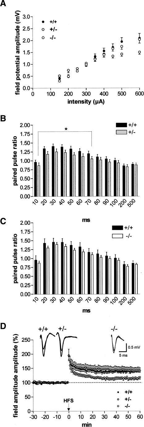
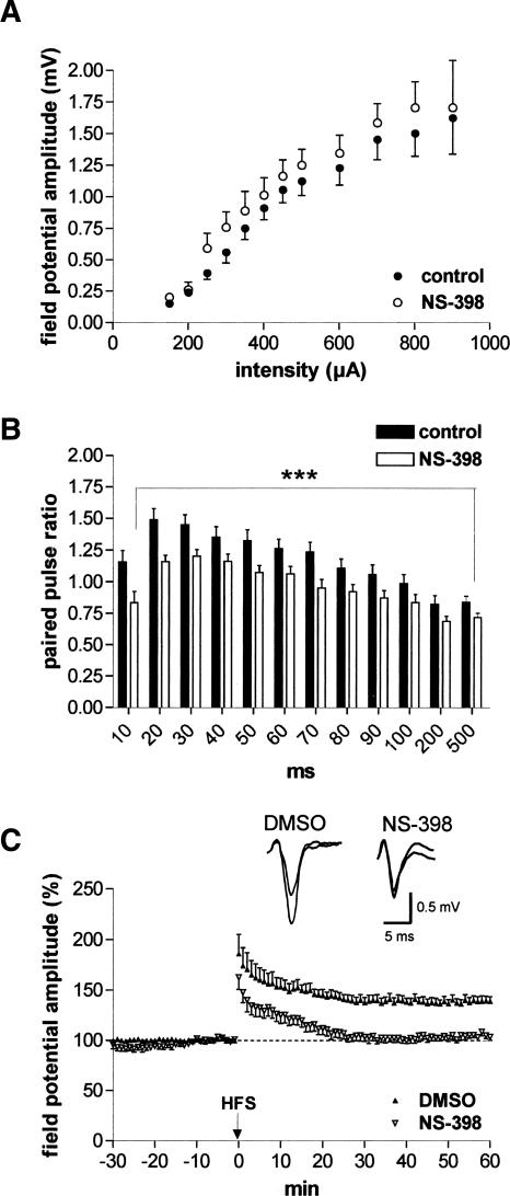
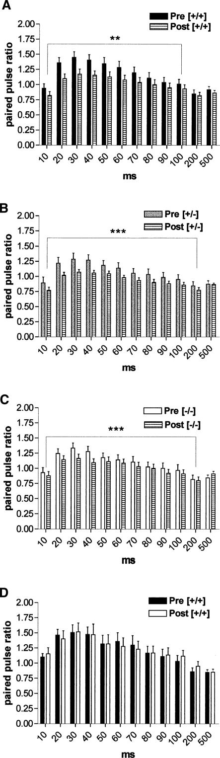
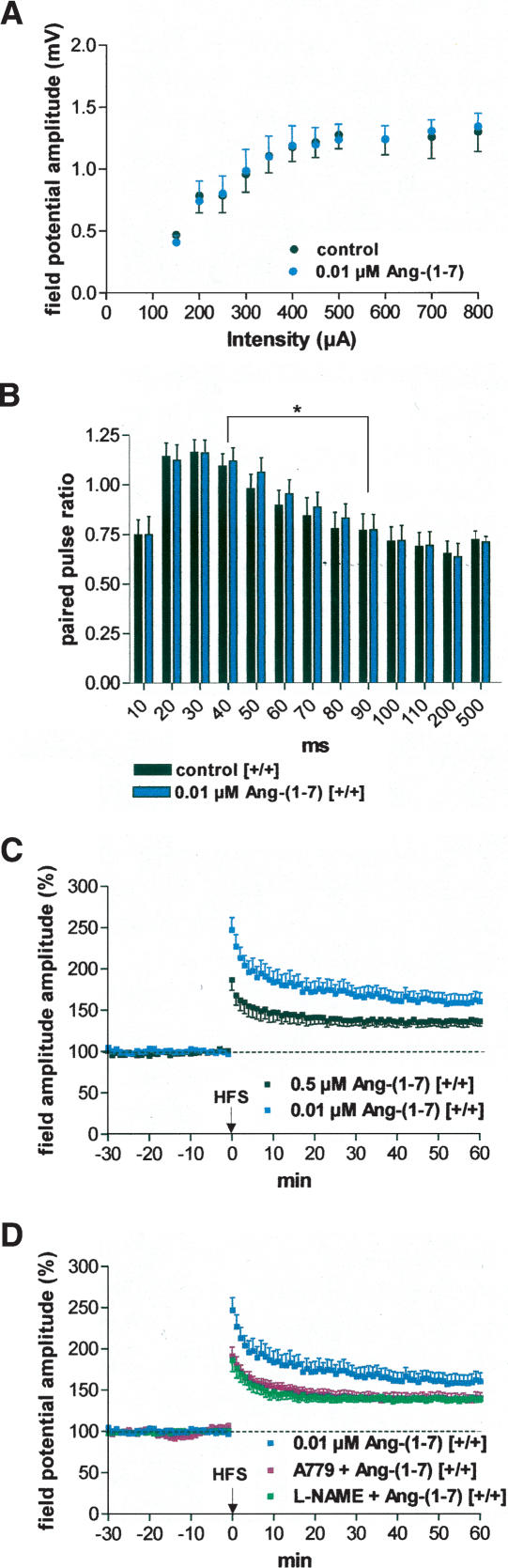
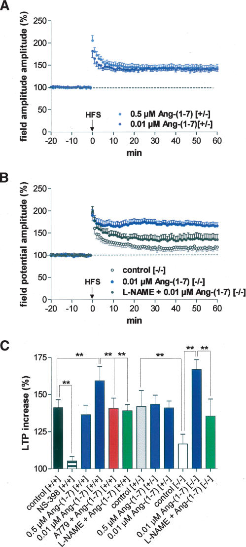
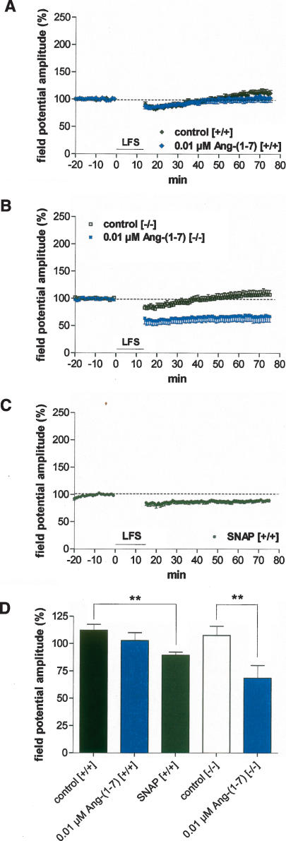
It is known from studies outside the brain that upon binding to its receptor, angiotensin-(1-7) elicits the release of prostanoids and nitric oxide (NO). Cyclooxygenase (COX) is a key enzyme that converts arachidonic acid to prostaglandins. Since there are no data available so far on the role of COX-2 in the amygdala, in a first step we demonstrated that the selective COX-2 inhibitor NS-398 significantly reduced the probability of long-term potentiation (LTP) induction in the lateral nucleus of the amygdala. Similarly, in COX-2(-/-) mice, LTP induced by external capsule (EC) stimulation was impaired. Second, we evaluated the action of angiotensin-(1-7) in the amygdala. In wild-type mice, angiotensin-(1-7) increased LTP. This LTP-enhancing effect of Ang-(1-7) was not observed in COX-2(+/-) mice. However, in COX-2(-/-) mice, Ang-(1-7) caused an enhancement of LTP similar to that in wild-type mice. The NO synthetase inhibitor L-NAME blocked this angiotensin-(1-7)-induced increase in LTP in COX-2(-/-) mice. Low-frequency stimulation of external capsule fibers did not cause long-term depression (LTD) in drug-free and angiotensin-(1-7)-treated brain slices in wild-type mice. In contrast, in COX-2(-/-) mice, angiotensin-(1-7) caused stable LTD. Increasing NO concentration by the NO-donor SNAP also caused LTD in wild-type mice. Our study shows for the first time that LTP in the amygdala is dependent on COX-2 activity. Moreover, COX-2 is involved in the mediation of angiotensin-(1-7) effects on LTP. Finally, it is recognized that there is a molecular cross-talk between COX-2 and NO that may regulate synaptic plasticity.


Figures

Figure 1.
Figure 1.
COX-2 influences excitability and plasticity in the lateral nucleus of the amygdala (LA). (A) Input/output curves, which demonstrate differences in field potential amplitudes between homozygous, heterozygous COX-2-deficient mice, and their wild-type littermates. Note a significant difference between wild-type and heterozygous COX-2-deficient mice at higher current intensities (400–600 μA, P < 0.03). (B) Paired-pulse ratios of field potential amplitudes in the LA in slices derived from (black bars) wild-type (+/+; n = 30) and (gray bars) heterozygous COX-2-deficient (+/−; n = 31) mice. At interstimulus intervals from 10 to 70 msec, a significant reduction of paired-pulse facilitation was found in heterozygous COX-2-deficient mice (P < 0.04). (C) Paired-pulse ratios of field potential amplitudes in the LA in slices derived from (black bars) wild-type (+/+; n = 18) and (white bars) homozygous COX-2-deficient (−/−; n = 39) mice. (D) High-frequency stimulation (HFS) of external capsule fibers elicited a reduced magnitude of LA-LTP in homozygous COX-2-deficient mice (−/−; n = 8) in comparison with heterozygous COX-2-deficient mice (+/−; n = 8) and their wild-type littermates (+/+; n = 9). Representative traces were recorded 5 min prior to tetanus and 60 min after tetanus in the lateral nucleus of the amygdala. Application of high-frequency stimulation (HFS; 2 × 100 Hz, duration 1 sec, interval 30 sec) at time 0. Data points represent averaged amplitudes (mean ± SEM) normalized with respect to baseline values.
Figure 2.
Figure 2.
Action of the selective COX-2 inhibitor NS-398 on excitability, paired-pulse facilitation, and LTP. (A) Input/output curves did not show significant changes between control recording and after NS-398 application (10 μM; n = 13). (B) NS-398 (10 μM) caused an impairment of paired-pulse facilitation (n = 13). (C) NS-398 (n = 7) significantly suppressed LA-LTP in wild-type mice in comparison to DMSO-treated controls (n = 7). Representative traces were recorded 5 min prior to tetanus and 60 min after tetanus in the lateral nucleus of the amygdala. Application of high-frequency stimulation (HFS; 2 × 100 Hz, duration 1 sec, interval 30 sec) at time 0. Data points represent averaged amplitudes (mean ± SEM) normalized with respect to baseline values.
Figure 3.
Figure 3.
Interaction between paired-pulse facilitation and LTP. (A) The plot of the ratios of second to first responses at varying interpulse intervals recorded 60 min after induction of LTP (Post) in wild-type mice indicates that responses in the LA were significantly less facilitated throughout interpulse intervals of 10–100 msec than control responses recorded before the application of HFS (Pre) (n = 21, P < 0.04). (B,C) Both in heterozygous (n = 25, P < 0.001) and homozygous (n = 21, P < 0.001) COX-2-deficient mice, a significant decrease in paired-pulse facilitation was obtained after induction of LTP over the range of interpulse intervals from 10 to 200 msec. (D) In slices in which NS-398 blocked the induction of LTP, paired-pulse facilitation recorded 60 min after tetanic stimulation did not differ from that recorded before washing in NS-398.
Figure 4.
Figure 4.
Dose-dependent effects of angiotensin-(1-7) on excitability and plasticity in the LA of slices derived from wild-type mice. (A) Ang-(1-7) (0.01 μM) did not change input/output curves significantly; (B) (blue bars) 0.01 μM Ang-(1-7) enhanced paired-pulse facilitation throughout interpulse intervals of 40–90 msec (n = 17); (C) 0.5 μM Ang-(1-7) (n = 8) did not change the magnitude of LA-LTP in comparison to drug-free controls (see Fig. 1D), whereas 0.01 μM significantly increased LA-LTP (n = 8). (D) A779 (0.01 μM), a specific Ang-(1-7) receptor antagonist, blocked the LTP-enhancing effect of Ang-(1-7) (n = 8). The unspecific inhibitor of the NO system L-NAME (200 μM) also blocked the Ang-(1-7)-mediated effect (n = 10). For comparison, the effect of 0.01 μM Ang-(1-7) on the induction of LTP is also presented. (C,D) Application of high-frequency stimulation (HFS; 2 × 100 Hz, duration 1 sec, interval 30 sec) at time 0. Data points represent averaged amplitudes (mean ± SEM) normalized with respect to baseline values.
Figure 5.
Figure 5.
Effects of Ang-(1-7) on LTP in COX-2-deficient mice. (A) In heterozygous COX-2-deficient mice (+/−), Ang-(1-7) neither at 0.5 μM (n = 7) nor at 0.01 μM (n = 7) was able to change the magnitude of LA-LTP in comparison to drug-free controls. (B) In homozygous COX-2-deficient mice (−/−), 0.01 μM Ang-(1-7) (n = 9) elicited a significant enhancement of LA-LTP. This enhancement of LTP could be blocked by 200 μM L-NAME (n = 7). For comparison, the control LTP recorded in COX-2−/− mice is also presented. (A,B) Application of high-frequency stimulation (HFS; 2 × 100 Hz, duration 1 sec, interval 30 sec) at time 0. Data points represent averaged amplitudes (mean ± SEM) normalized with respect to baseline values. (C) Bar histogram of data points shown in Figures 3 and 4, as averaged 55–60 min after HFS and normalized with respect to baseline (mean ± SEM). Significant differences are indicated; (**) P < 0.001.
Figure 6.
Figure 6.
Effects of 0.01 μM Ang-(1-7) on low-frequency stimulation (LFS) induced activity change in the LA. (A) LFS caused a significant long-term depression (LTD) neither in drug-free controls (n = 6) nor in Ang-(1-7)-treated slices (n = 7) derived from wild-type mice (+/+). (B) In homozygous COX-2-deficient mice (−/−), 0.01 μM Ang-(1-7) elicited a significant, long-lasting reduction of field potential amplitudes (n = 5). In contrast, no LTD was observed in drug-free homozygous COX-2-deficient slices (n = 7). (C) SNAP (NO donor, 100 μM) induced a weak LTD in wild-type mice. (D) Bar histogram of data points shown in Figure 5A–C, as averaged 55–60 min after LFS and normalized with respect to baseline (mean ± SEM). Significant differences are indicated; (**) P < 0.001.

References

    1. Albrecht D., Nitschke T., von Bohlen und Halbach O. Various effects of angiotensin II on amygdaloid neuronal activity in normotensive control and hypertensive transgenic [TGR(mREN-2)27] rats. FASEB J. 2000;14:925–931. - PubMed
    1. Andreasson K.I., Savonenko A., Vidensky S., Goellner J.J., Zhang Y., Shaffer A., Kaufmann W.E., Worley P.F., Isakson P., Markowska A.L. Age-dependent cognitive deficits and neuronal apoptosis in cyclooxygenase-2 transgenic mice. J. Neurosci. 2001;21:8198–8209. - PMC - PubMed
    1. Arima S., Ito S. New insights into actions of the renin-angiotensin system in the kidney: Concentrating on the Ang II receptors and the newly described Ang-(1-7) and its receptor. Semin. Nephrol. 2001;21:535–543. - PubMed
    1. Armstrong D.L., Garcia E.A., Ma T., Quinones B., Wayner M.J. Angiotensin-II blockade of long-term potentiation at the perforant path-granule cell synapse in-vitro. Peptides. 1996;17:689–693. - PubMed
    1. Bidmon H.J., Oermann E., Schiene K., Schmitt M., Kato K., Asayama K., Witte O.W., Zilles K. Unilateral upregulation of cyclooxygenase-2 following cerebral, cortical photothrombosis in the rat: Suppression by MK-801 and co-distribution with enzymes involved in the oxidative stress cascade. J. Chem. Neuroanat. 2000;20:163–176. - PubMed

MeSH terms

LinkOut - more resources