. 2007 Apr;3(4):222-8.
doi: 10.1038/nchembio868. Epub 2007 Mar 11.

Fluorogenic probes for monitoring peptide binding to class II MHC proteins in living cells

Affiliations

Fluorogenic probes for monitoring peptide binding to class II MHC proteins in living cells

Prasanna Venkatraman et al. Nat Chem Biol. 2007 Apr.

Abstract

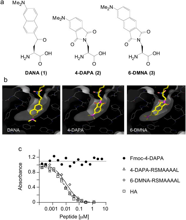
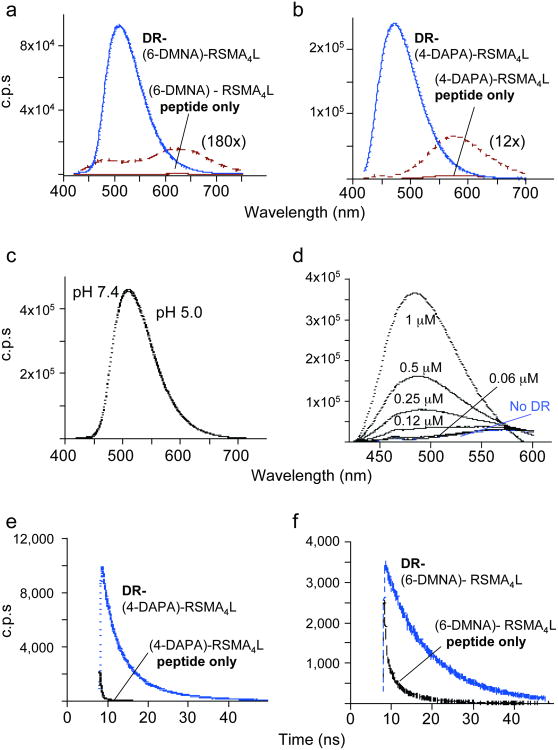
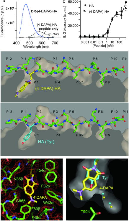
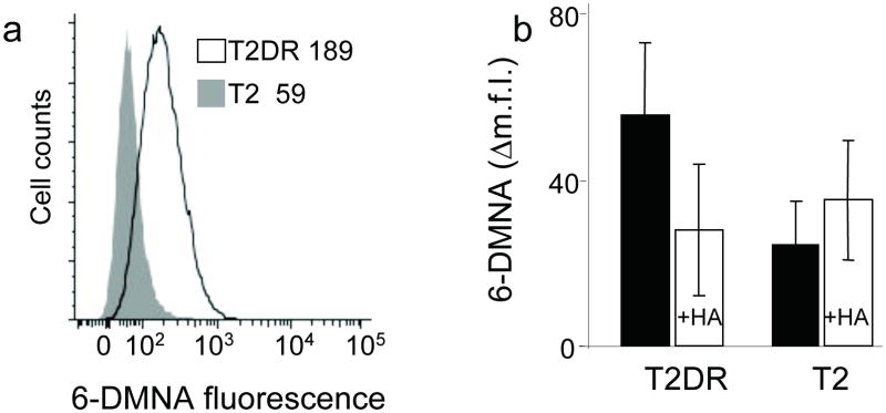
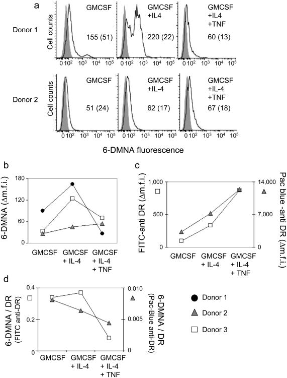
A crucial step in the immune response is the binding of antigenic peptides to major histocompatibility complex (MHC) proteins. Class II MHC proteins present their bound peptides to CD4(+) T cells, thereby helping to activate both the humoral and the cellular arms of the adaptive immune response. Peptide loading onto class II MHC proteins is regulated temporally, spatially and developmentally in antigen-presenting cells. To help visualize these processes, we have developed a series of novel fluorogenic probes that incorporate the environment-sensitive amino acid analogs 6-N,N-dimethylamino-2-3-naphthalimidoalanine and 4-N,N-dimethylaminophthalimidoalanine. Upon binding to class II MHC proteins these fluorophores show large changes in emission spectra, quantum yield and fluorescence lifetime. Peptides incorporating these fluorophores bind specifically to class II MHC proteins on antigen-presenting cells and can be used to follow peptide binding in vivo. Using these probes we have tracked a developmentally regulated cell-surface peptide-binding activity in primary human monocyte-derived dendritic cells.


Figures

Figure 1
Figure 1. 4-DAPA and 6-DMNA can be modeled into the P1 pocket of DR1 without major distortion
(a) Chemical structures of DANA, 4-DAPA and 6-DMNA. (b) In the crystal structure of the DR1-HA peptide complex, a tyrosine residue is buried deep into the hydrophobic P1 pocket . DANA, 4-DAPA and 6-DMNA were modeled in silico into this pocket in place of tyrosine. Residues shown lining the pocket are (clockwise from upper right) Pheα54, Pheα32, Trpα43 (behind), Ileα7, Trpβ153, Pheα48, Thrβ90, Valβ91, Tyrβ83, Glyβ86, and Valβ85. The side chains of Asnβ82 and Hisβ81 (upper left) form hydrogen bonds with the peptide main chain at the mouth of the P1 pocket. Other residues lining the pocket but not shown are Pheα24, Pheα26, and Pheα48 (c) Binding of fluorogenic peptides to DR1 assessed using a competitive binding assay. DR1 was incubated with biotin-HA peptide and various concentrations of unlabelled inhibitor peptides, with biotin-HA binding quantified by a sandwich ELISA assay using streptavidin alkaline phosphatase. Binding of Fmoc-(4-DAPA) was assessed to evaluate non-specific binding of the fluorophore. IC50 values for these and other peptides (Supplementary Table I online) were determined as described (Supplementary Methods online).
Figure 2
Figure 2. Spectral properties of 6-DMNA and 4-DAPA peptides free in solution and bound to DR1
Fluorescence emission spectra of (a) (6-DMNA)-RSMA4L (20 nM) and (b) (4-DAPA)-RSMA4L (400 nM) peptides and their complexes with HLA-DR1 are shown, using 400 nm excitation. Free peptide spectra are shown also on an expanded scale. Note the shift in emission λmax upon binding of the peptide to DR and the increase in the emission intensity. Fluorescence intensities are reported as counts per second (c.p.s.). (c) The fluorescence spectrum of the purified DR-(6-DMNA) complex was recorded in citrate phosphate buffer (pH 5.0) and in PBS (pH 7.4). No difference was observed between the two pH. (d) Various concentrations of DR1 were incubated with 1 μM (4-DAPA)-RSMA4L and the emission spectra of the mixtures recorded. (f,g) Fluorescence lifetime measurements. Fluorescence lifetime is increased upon binding of the (4-DAPA)-RSMA4L (f) and (6-DMNA)-RSMA4L (g) to DR. Fluorescence lifetime values measured by time-correlation single-photon counting spectroscopy (Supplementary Table 3 online) were determined as described (Supplementary Methods online).
Figure 3
Figure 3. Structural and functional characterization of (4-DAPA)-HA peptide
(a) (4-DAPA)-HA was incubated with HLA-DR1 as described for (4-DAPA)-RSMA4L, and the complex formed was purified by gel filtration. Fluorescence of DR-(4-DAPA)-HA was compared with the free peptide. (b) Activation of HA1.7 TCR hybridoma by antigen presenting cells pulsed with HA (filled circles) or (4-DAPA)-HA (closed triangles) peptides. T cell activation reported as counts per minute (c.p.m.) measured in a thymidine incorporation bioassay for IL-2 as secreted by activated T cells. Error bars indicated standard deviation of triplicate measurements. (c-e) Crystal structure of (4-DAPA)-HA bound to DR1. (c) top, (4-DAPA)-HA peptide shown with surface representation of the DR1 peptide binding site, with 4-DAPA side chain shown with yellow bonds extending down into the P1 pocket; bottom, unmodified HA peptide from the crystal structure of the DR1-HA-SEC (3B2) complex (PDB ID 1JWU) shown after alignment of MHC peptide binding domain, with tyrosine side chain at the P1 position shown with magenta bonds. (d) ∣2Fo−Fc∣ omit map of the region around the P1 pocket with all residues shown removed from the model before map calculation. (e) section through the P1 pocket, showing the HA peptide tyrosine side chain and the (4-DAPA)-HA fluorophore, along with the corresponding ordered water molecules, colored as in (c). Panels (c-f) made using Pymol.
Figure 4
Figure 4. (6-DMNA)-peptide binds specifically to DR molecules expressed on live cells
(a) T2 cells expressing HLA-DR1 (T2DR, dark line) or untransfected T2 cells (shaded profile) were incubated with (6-DMNA)-RSMA4L under conditions which inhibit endocytosis, and the fluorescence of the bound peptide was measured by flow cytometry. Numbers in inset indicate the mean fluorescence intensity values for the indicated cells. (b) Binding of (6-DMNA)-RSMA4L peptide to HLA-DR1 on these cells was reduced in the presence of excess unlabelled HA peptide. The Δm.f.i. values shown are mean fluorescence intensity of labeled cells minus background autofluorescence from the same cells, and represent the average and standard deviation of three independent experiments.
Figure 5
Figure 5. (6-DMNA)-peptide binds to human monocyte-derived DC in a maturation-dependent manner
Peptide binding to monocyte derived DC subsets isolated from HLA-DR1+ donors and prepared by an in vitro differentiation protocol (see methods) was performed essentially as described for T2DR. (a) Fluorescent peptide binding to DC subsets isolated from two different donors. Solid line, (6-DMNA)-RSMA4L, shaded curve, autofluorescence. Numbers indicate the mean fluorescence intensity for peptide treated cells (with autofluorescence intensity for the same cells shown in parenthesis). (b) Δm.f.i. values for DC subsets generated from cells obtained from three different donors. (c) Expression of HLA-DR1 was measured using murine anti human HLA-DR monoclonal antibody L243 labeled with FITC or Pacific-Blue, for DC subsets from two of the donors in (b). (d) Peptide binding expressed as the ratio of 6-DMNA fluorescence to FITC-anti-DR or Pacific-Blue-anti-DR antibody fluorescence.

Comment in

References

    1. Trombetta ES, Mellman I. Cell biology of antigen processing in vitro and in vivo. Annu Rev Immunol. 2005;23:975–1028. - PubMed
    1. McFarland BJ, Beeson C. Binding interactions between peptides and proteins of the class II major histocompatibility complex. Med Res Rev. 2002;22:168–203. - PubMed
    1. Jardetzky TS, et al. Peptide binding to HLA-DR1: a peptide with most residues substituted to alanine retains MHC binding. Embo J. 1990;9:1797–1803. - PMC - PubMed
    1. Sato AK, et al. Determinants of the peptide-induced conformational change in the human class II major histocompatibility complex protein HLA-DR1. J Biol Chem. 2000;275:2165–2173. - PubMed
    1. Stern LJ, et al. Crystal structure of the human class II MHC protein HLA-DR1 complexed with an influenza virus peptide. Nature. 1994;368:215–221. - PubMed

Publication types

MeSH terms