

Pancreatic carcinoma coexisting with chronic pancreatitis versus tumor-forming pancreatitis: diagnostic utility of the time-signal intensity curve from dynamic contrast-enhanced MR imaging
- PMID: 17352014
- PMCID: PMC4065920
- DOI: 10.3748/wjg.v13.i6.858
Pancreatic carcinoma coexisting with chronic pancreatitis versus tumor-forming pancreatitis: diagnostic utility of the time-signal intensity curve from dynamic contrast-enhanced MR imaging
Abstract
Aim: To evaluate the ability of the time-signal intensity curve (TIC) of the pancreas obtained from dynamic contrast-enhanced magnetic resonance imaging (MRI) for differentiation of focal pancreatic masses, especially pancreatic carcinoma coexisting with chronic pancreatitis and tumor-forming pancreatitis.
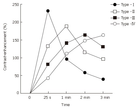
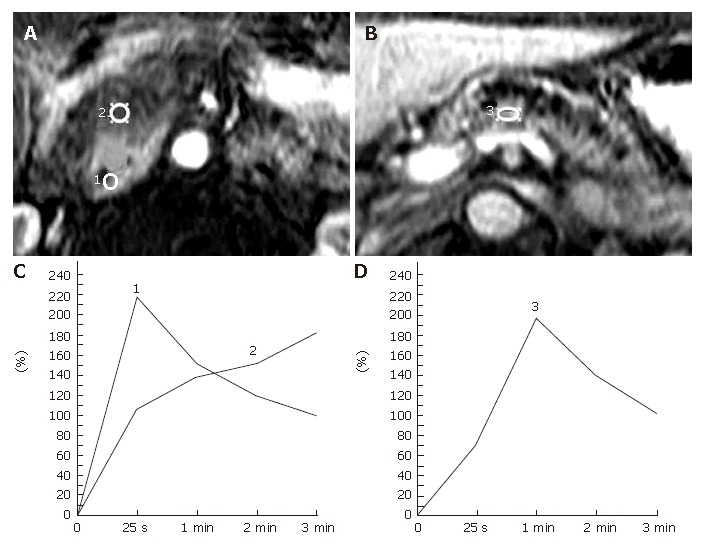
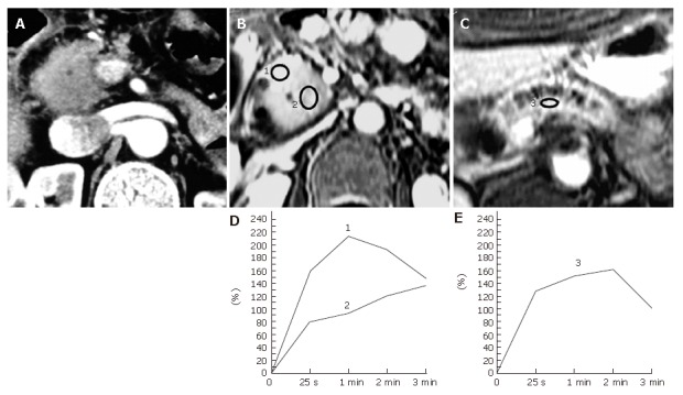
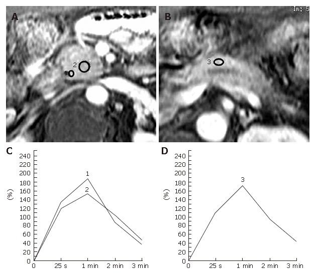
Methods: Forty-eight consecutive patients who underwent surgery for a focal pancreatic mass, including pancreatic ductal carcinoma (n=33), tumor-forming pancreatitis (n=8), and islet cell tumor (n=7), were reviewed. Five pancreatic carcinomas coexisted with longstanding chronic pancreatitis. The pancreatic TICs were obtained from the pancreatic mass and the pancreatic parenchyma both proximal and distal to the mass lesion in each patient, prior to surgery, and were classified into 4 types according to the time to a peak: 25 s and 1, 2, and 3 min after the bolus injection of contrast material, namely, type-I, II, III, and IV, respectively, and were then compared to the corresponding histological pancreatic conditions.
Results: Pancreatic carcinomas demonstrated type-III (n=13) or IV (n=20) TIC. Tumor-forming pancreatitis showed type-II (n=5) or III (n=3) TIC. All islet cell tumors revealed type-I. The type-IV TIC was only recognized in pancreatic carcinoma, and the TIC of carcinoma always depicted the slowest rise to a peak among the 3 pancreatic TICs measured in each patient, even in patients with chronic pancreatitis.
Conclusion: Pancreatic TIC from dynamic MRI provides reliable information for distinguishing pancreatic carcinoma from other pancreatic masses, and may enable us to avoid unnecessary pancreatic surgery and delays in making a correct diagnosis of pancreatic carcinoma, especially, in patients with longstanding chronic pancreatitis.
Figures
References
-
- Steer ML, Waxman I, Freedman S. Chronic pancreatitis. N Engl J Med. 1995;332:1482–1490. - PubMed
-
- Johnson PT, Outwater EK. Pancreatic carcinoma versus chronic pancreatitis: dynamic MR imaging. Radiology. 1999;212:213–218. - PubMed
-
- Kim T, Murakami T, Takamura M, Hori M, Takahashi S, Nakamori S, Sakon M, Tanji Y, Wakasa K, Nakamura H. Pancreatic mass due to chronic pancreatitis: correlation of CT and MR imaging features with pathologic findings. AJR Am J Roentgenol. 2001;177:367–371. - PubMed
-
- Lowenfels AB, Maisonneuve P, Cavallini G, Ammann RW, Lankisch PG, Andersen JR, Dimagno EP, Andrén-Sandberg A, Domellöf L. Pancreatitis and the risk of pancreatic cancer. International Pancreatitis Study Group. N Engl J Med. 1993;328:1433–1437. - PubMed
Publication types
MeSH terms
LinkOut - more resources
Full Text Sources
Medical
