. 2007 Feb 14;13(6):874-81.
doi: 10.3748/wjg.v13.i6.874.

Effects of hypothalamic paraventricular nuclei on apoptosis and proliferation of gastric mucosal cells induced by ischemia/reperfusion in rats

Affiliations

Effects of hypothalamic paraventricular nuclei on apoptosis and proliferation of gastric mucosal cells induced by ischemia/reperfusion in rats

Li Li et al. World J Gastroenterol. .

Abstract

Aim: To investigate the effects of electrical stimulation of hypothalamic paraventricular nuclei (PVN) on gastric mucosal cellular apoptosis and proliferation induced by gastric ischemia/reperfusion (I/R) injury.

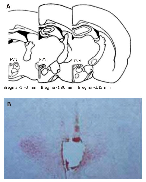
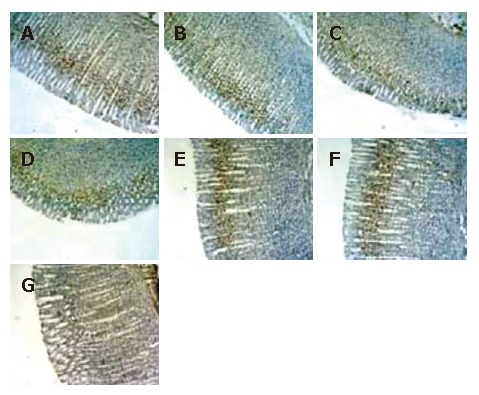
Methods: For different experimental purposes, stimulating electrode plantation or electrolytic destruction of the PVN was applied, then the animals' GI/R injury model was established by clamping the celiac artery for 30 min and allowing reperfusing the artery for 30 min, 1 h, 3 h or 6 h respectively. Then histological, immunohistochemistry methods were used to assess the gastric mucosal damage index, the gastric mucosal cellular apoptosis and proliferation at different times.

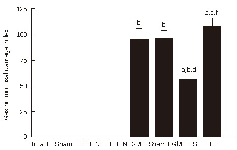
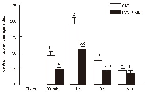
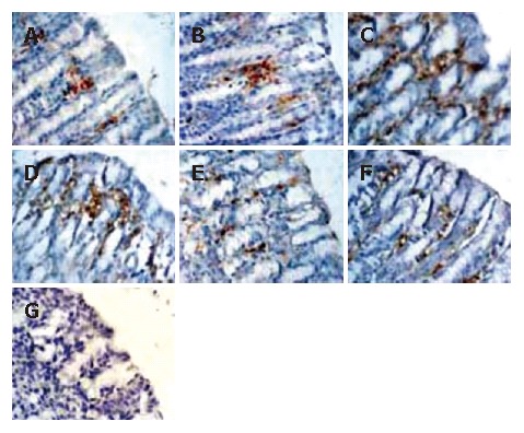
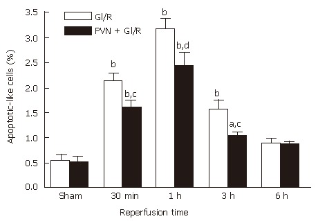
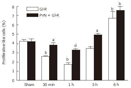
Results: The electrical stimulation of PVN significantly attenuated the GI/R injury at 30 min, 1 h and 3 h after reperfusion. The electrical stimulation of PVN decreased gastric mucosal apoptosis and increased gastric mucosal proliferation. The electrolytic destruction of the PVN could eliminate the protective effects of electrical stimulation of PVN on GI/R injury. These results indicated that the PVN participated in the regulation of GI/R injury as a specific area in the brain, exerting protective effects against the GI/R injury, and the protection was associated with the inhibition of cellular apoptosis and the promotion of gastric mucosal proliferation.

Conclusion: Stimulating PVN significantly inhibits the gastric mucosal cellular apoptosis and promots gastric mucosal cellular proliferation. This may explain the protective mechanisms of electrical stimulation of PVN against GI/R injury.


Figures

Figure 1
Figure 1
Sites of the stimulating electrode tip in PVN. A: Standard atlas sections of the rat brain showing the distributions of stimulating sites of the experimental animals; B: Photomicrographs of stimulating sites in the rat brain. The section stained with neutral red, showing the position of stimulating electrode tip by passing a positive DC of 1 mA for 10 s, which indicates a placement within the PVN.
Figure 2
Figure 2
Effect of electrical stimulation and bilateral electrolytic destruction of PVN on gastric mucosal damage induced by I/R at 1 h after reperfusion in rats. Intact: normal rat; sham: sham-operation (electrode was inserted, with no current passing and no artery clamped); ES + N: intact stomach after electrical stimulation of the PVN. EL + N: intact stomach after bilateral electrolytic destruction of the PVN; GI/R injury: gastric ischemia-reperfusion injury; Sham+GI/R injury: sham electrical stimulation of the PVN plus GI/R injury; ES: electrical stimulation of the PVN plus GI/R injury; EL: bilateral electrolytic destruction of the PVN plus GI/R injury. Values of GMDI are mean ± SE (n = 6). bP < 0.01 vs intact, dP < 0.01 ES vs ES + N, fP < 0.01 EL vs EL + N, aP < 0.05, ES vs GI-R, cP < 0.05, EL vs ES.
Figure 3
Figure 3
Effect of electrical stimulation of PVN on gastric mucosal damage induced by I/R at different times in rats. Sham: sham-operation(electrode was inserted, with no current passing and no artery clamped); GI/R: gastric ischemia/reperfusion was maintained for 30 min, 1 h, 3 h and 6 h after 30 min of ischemia, respectively; PVN + GI/R: electrical stimulation of the PVN plus GI/R. Values: Each column represents mean ± SE (n = 6). bP < 0.01 vs sham; aP < 0.05, dP < 0.01, PVN + GI/R vs GI/R at different times.
Figure 4
Figure 4
Histological exhibition of electrical stimulation of PVN on gastric mucosal apoptosis induced by I/R at different times in rats. The apoptotic-positive cells were probed with anti-M30 CytoDEATH antibody and counter-stained with hematoxylin in rat gastric mucosa (× 400). A: normal gastric mucosa; B: sham operation; C: GI/R at 1 h after reperfusion; D: PVN + GI/R at 1 h after reperfusion; E: GI/R at 6 h after reperfusion; F: PVN+GI/R at 6 h after reperfusion; G: negative control group.
Figure 5
Figure 5
Effect of electrical stimulation of PVN on gastric mucosal cellular apoptosis induced by I/R at different time points in rats. Sham: sham-operation; GI/R: reperfusion was maintained for 30 min, 1 h, 3 h and 6 h after 30 min of ischemia, respectively; PVN + GI/R: electrical stimulation of PVN plus GI/R. The percentage of apoptotic cells was taken by counting the cells in 10 microscopic fields (× 400). Each column represents mean ± SE (n = 6). aP < 0.05, bP < 0.01, GI/R vs sham-operation; cP < 0.05, dP < 0.01, PVN + GI/R vs GI/R at each time point.
Figure 6
Figure 6
Histological exhibition of electrical stimulation of PVN on gastric mucosal cellular proliferation induced by I/R at different times in rats. Proliferating-positive cells were probed with anti-mouse anti-proliferating cell nuclear antigen (PCNA) antibody and counter stained with hematoxylin in gastric mucosa (× 100). A: normal control gastric mucosa; B: gastric mucosa after sham operation; C: GI/R, 1 h after reperfusion; D: PVN + GI/R, 1 h after reperfusion; E: GI/R, 6 h after reperfusion; F: PVN + GI-R, 6 h after reperfusion; G: negative control group.
Figure 7
Figure 7
Effect of electrical stimulation of PVN on gastric mucosal proliferation induced by I/R at different times in rats. Sham: sham-operation; GI/R: reperfusion was maintained for 30 min, 1 h, 3 h and 6 h after 30 min of ischemia, respectively; PVN+GI/R: electrical stimulation of PVN plus GI/R. The percentage of proliferating cells was taken by using cell count in 10 microscopic fields (× 100). Each column represents an average value expressed in mean ± SE (n = 6). bP < 0.01, GI/R vs sham operation; aP < 0.05 and dP < 0.01, PVN + GI/R vs GI/R.

Similar articles

Cited by

References

    1. De La Lastra CA, Cabeza J, Motilva V, Martin MJ. Melatonin protects against gastric ischemia-reperfusion injury in rats. J Pineal Res. 1997;23:47–52. - PubMed
    1. Kishimoto Y, Wada K, Nakamoto K, Ashida K, Kamisaki Y, Kawasaki H, Itoh T. Quantitative analysis of cyclooxygenase-2 gene expression on acute gastric injury induced by ischemia-reperfusion in rats. Life Sci. 1997;60:PL127–PL133. - PubMed
    1. Kitano M, Wada K, Kamisaki Y, Nakamoto K, Kishimoto Y, Kawasaki H, Itoh T. Effects of cimetidine on acute gastric mucosal injury induced by ischemia-reperfusion in rats. Pharmacology. 1997;55:154–164. - PubMed
    1. Wada K, Kamisaki Y, Kitano M, Nakamoto K, Itoh T. Protective effect of cystathionine on acute gastric mucosal injury induced by ischemia-reperfusion in rats. Eur J Pharmacol. 1995;294:377–382. - PubMed
    1. Andrews FJ, Malcontenti-Wilson C, O'Brien PE. Polymorphonuclear leukocyte infiltration into gastric mucosa after ischemia-reperfusion. Am J Physiol. 1994;266:G48–G54. - PubMed

Publication types