. 2007 Apr 3;46(13):3942-51.
doi: 10.1021/bi700130e. Epub 2007 Mar 13.

3'-Phosphoadenosine-5'-phosphosulfate reductase in complex with thioredoxin: a structural snapshot in the catalytic cycle

Affiliations

3'-Phosphoadenosine-5'-phosphosulfate reductase in complex with thioredoxin: a structural snapshot in the catalytic cycle

Justin Chartron et al. Biochemistry. .

Abstract

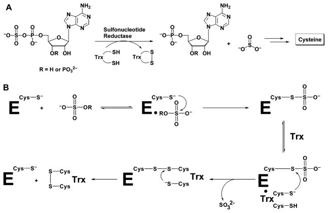
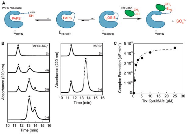
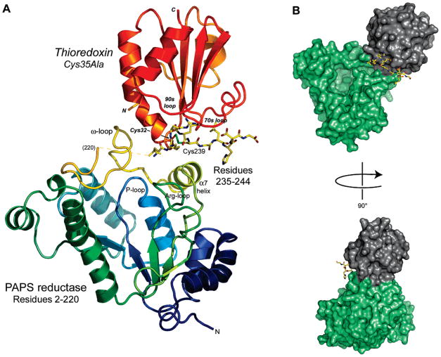
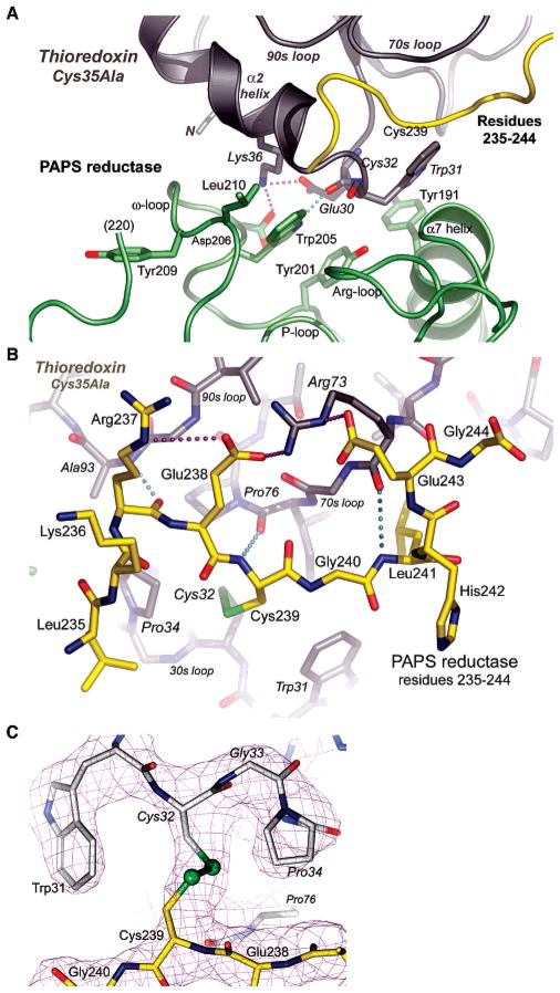
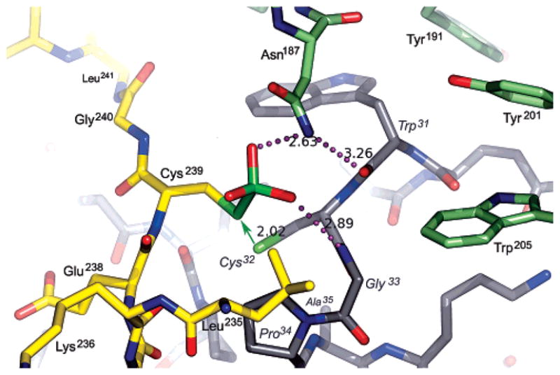
The crystal structure of Escherichia coli 3'-phosphoadenosine-5'-phosphosulfate (PAPS) reductase in complex with E. coli thioredoxin 1 (Trx1) has been determined to 3.0 A resolution. The two proteins are covalently linked via a mixed disulfide that forms during nucleophilic attack of Trx's N-terminal cysteine on the Sgamma atom of the PAPS reductase S-sulfocysteine (E-Cys-Sgamma-SO3-), a central intermediate in the catalytic cycle. For the first time in a crystal structure, residues 235-244 in the PAPS reductase C-terminus are observed, depicting an array of interprotein salt bridges between Trx and the strictly conserved glutathione-like sequence, Glu238Cys239Gly240Leu241His242. The structure also reveals a Trx-binding surface adjacent to the active site cleft and regions of PAPS reductase associated with conformational change. Interaction at this site strategically positions Trx to bind the S-sulfated C-terminus and addresses the mechanism for requisite structural rearrangement of this domain. An apparent sulfite-binding pocket at the protein-protein interface explicitly orients the S-sulfocysteine Sgamma atom for nucleophilic attack in a subsequent step. Taken together, the structure of PAPS reductase in complex with Trx highlights the large structural rearrangement required to accomplish sulfonucleotide reduction and suggests a role for Trx in catalysis beyond the paradigm of disulfide reduction.


Figures

Figure 1
Figure 1
(A) Depending on the organism, APS or PAPS is reduced to sulfite by APS reductase or PAPS reductase, respectively. The sulfite product formed in this reaction is further reduced to sulfide, and the sulfur is incorporated into cysteine. This amino acid is then converted into numerous metabolites, including methionine and cofactors, such as coenzyme A. (B) Mechanism proposed for sulfonucleotide reduction.
Figure 2
Figure 2
Formation of the PAPS reductase–Trx1 protein complex. (A) Strategy for trapping the E. coli PAPS reductase–Trx1 protein complex. (B) Complex formation was monitored at 220 nm by reversed phase HPLC analysis and plotted as a function of time. E. coli Trx1 Cys35Ala (10 μM) was incubated with increasing amounts PAPS reductase S-sulfocysteine intermediate [0 (i), 4 (ii), 9 (iii), and 20 μM (iv) at the left] or PAPS reductase alone [0 (i) and 20 μM (iv) at the right]. Mass spectrometry analysis of the observed peaks indicated masses of 13 937.1 Da for Trx1 [(●) 13 937.8 Da, theoretical], 30 007.3 Da for PAPS reductase [(■) 30 007.5 Da, theoretical], and 43 941.8 Da for the PAPS reductase–Trx1 complex [(*) 43 942.4 Da, theoretical]. (C) Rate of formation of the PAPS reductase–Trx1 complex from the PAPS reductase S-sulfocysteine intermediate (1 μM) plotted as a function of Trx Cys35Ala concentration, monitored by SYPRO ruby-stained nonreducing SDS–PAGE gel analysis. The dashed line represents a fit of the data to the equation derived for active site-directed irreversible inhibitors as described in Experimental Procedures and gave an apparent Ki value of 1.1 μM for the PAPS reductase S-sulfocysteine intermediate and Trx1 Cys35Ala.
Figure 3
Figure 3
Overall view of the PAPS reductase–Trx1 complex in the cocrystal structure. (A) The flexible C-terminal tail of the reductase (residues 235–244; all atoms shown) fits into a groove on Trx1 comprised of the 30s, 70s, and 90s loops (Trx1 residues 2833, 7177, 9196, respectively; Trx1 residues in italics); the mixed disulfide is formed between Cys239 of the reductase and Cys32 of Trx. Trx in turn is bound to PAPS reductase among an ω-loop (residues 202–212, colored yellow), helix α7, and the C-terminal Cys239-peptide (residues 235–244). Residues 221–234 of PAPS reductase are disordered in the cocrystal structure (indicated by a dashed yellow line in front of the ω-loop). (B) Solvent accessible surface depiction of the PAPS reductase–Trx1 complex. In panel B and in Figures 4 and 5, PAPS reductase residues 1–220 are colored green, C-terminal peptide residues 235–244 are colored yellow, and Trx is colored gray.
Figure 4
Figure 4
(A) Specific recognition of PAPS reductase by Trx1 occurs via residues in an ω-loop of the reductase and the 30s loop of Trx1 and involves hydrogen bonds, aromatic stacking, and hydrophobic interactions. The view is the same as in Figure 3A. (B) Specific recognition of the C-terminal peptide of PAPS reductase (yellow) by Trx1 (gray) involves three hydrogen bonds between main chain atoms, hydrophobic contacts with Leu235 in a pocket on TrxA, and interdigitation of Arg237, Glu238, Arg73, and Glu243 in a network of three salt bridges. The view is the same as in Figure 3A. (C) Electron density for the mixed disulfide formed between PAPS reductase Cys239 and Trx1 Cys32 (σA-weighted 2|Fo| − |Fc| map contoured at 1.7σ). Density for adjacent residues in the PAPS reductase C-terminal peptide (yellow) and in the TrxA 30s loop (gray) is also shown. The view is from behind the complex as shown in Figure 3A (i.e., the Glu238Cys239Gly240 peptide orientation is reversed).
Figure 5
Figure 5
Model for the S-sulfocysteine intermediate of PAPS reductase bound to Trx1, based on the crystal structure of the mixed disulfide complex. The sulfite moiety fits into a pocket at the PAPS reductase–Trx1 interface. PAPS reductase residues in the C-terminal tail are colored yellow and other residues green; Trx1 residues are colored gray with labels in italics. Potential hydrogen bonds (dotted lines; distances in angstroms) include those from Asn187 to the sulfite and the carbonyl of Trp31 and from the amide of Gly33 to the sulfite. The green arrow represents the nucleophilic attack that must occur for formation of the mixed disulfide and displacement of sulfite; the distance between the Sγ atoms of Cys239 and Cys32 in the cocrystal structure is 2.02 Å. The resolving cysteine of Trx, Cys35, is mutated to Ala in the complex (visible below Pro34 in the 30s loop). With respect to Figure 3A, the view is approximately from the N-terminus of Trx1 looking along the axis of the C-terminal peptide.

References

    1. Kredich NM. Escherichia coli and Salmonella typhimurium: Cellular and Molecular Biology. Vol. 1. American Society for Microbiology; Washington, DC: 1996.
    1. Schwenn JD. Photosynthetic Sulphate Reduction. Z Naturforsch. 1994;49c:531–539.
    1. Carroll KS, Gao H, Chen H, Stout CD, Leary JA, Bertozzi CR. A conserved mechanism for sulfonucleotide reduction. PLoS Biol. 2005;3:e250. - PMC - PubMed
    1. Kopriva S, Buchert T, Fritz G, Suter M, Weber M, Benda R, Schaller J, Feller U, Schurmann P, Schunemann V, Trautwein AX, Kroneck PM, Brunold C. Plant adenosine 5′-phosphosulfate reductase is a novel iron-sulfur protein. J Biol Chem. 2001;276:42881–42886. - PubMed
    1. Bick JA, Dennis JJ, Zylstra GJ, Nowack J, Leustek T. Identification of a new class of 5′-adenylylsulfate (APS) reductases from sulfate-assimilating bacteria. J Bacteriol. 2000;182:135–142. - PMC - PubMed

Publication types

Substances

Associated data

LinkOut - more resources