. 2007 Mar;3(3):e31.
doi: 10.1371/journal.ppat.0030031.

Conversion of the BASE prion strain into the BSE strain: the origin of BSE?

Affiliations

Conversion of the BASE prion strain into the BSE strain: the origin of BSE?

Raffaella Capobianco et al. PLoS Pathog. 2007 Mar.

Abstract

Atypical neuropathological and molecular phenotypes of bovine spongiform encephalopathy (BSE) have recently been identified in different countries. One of these phenotypes, named bovine "amyloidotic" spongiform encephalopathy (BASE), differs from classical BSE for the occurrence of a distinct type of the disease-associated prion protein (PrP), termed PrP(Sc), and the presence of PrP amyloid plaques. Here, we show that the agents responsible for BSE and BASE possess different biological properties upon transmission to transgenic mice expressing bovine PrP and inbred lines of nontransgenic mice. Strikingly, serial passages of the BASE strain to nontransgenic mice induced a neuropathological and molecular disease phenotype indistinguishable from that of BSE-infected mice. The existence of more than one agent associated with prion disease in cattle and the ability of the BASE strain to convert into the BSE strain may have important implications with respect to the origin of BSE and spongiform encephalopathies in other species, including humans.


Conflict of interest statement

Competing interests. The authors have declared that no competing interests exist.

Figures

Figure 1
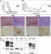
Figure 1. Primary Transmission of BSE and BASE to Tgbov XV Mice
(A) Survival curves and (B) lesion profiles for mice infected with BSE and BASE. (B) Vacuolation was scored on a scale of 0–5 in the following brain areas: 1, dorsal medulla; 2, cerebellar cortex; 3, superior colliculus; 4, hypothalamus; 5, thalamus; 6, hippocampus; 7 septum; 8, retrosplenial and adjacent motor cortex; and 9, cingulate and adjacent motor cortex. Data are mean ± s.e.m. (C–J) Neuropathological changes in mice infected with BSE (C–F) and BASE (G–J). Micrographs were obtained from corresponding areas of the hippocampal region and cerebral cortex stained with haematoxylin-eosin (C, D, G, and H), or labeled with the anti-PrP antibody 6H4 (E, F, I, and J). The neuropathological profile of BSE-infected mice is marked by the presence of numerous amyloid plaques while spongiform changes are mild (C and D). Conversely, mice challenged with BASE show a severe vacuolation in the absence of amyloid deposits (G and H). PrP immunohistochemistry shows uni- and multicentric amyloid plaques associated with granular deposits in BSE-infected mice (E and F) and diffuse immunostaining of the neuropil with focal enhancement in mice challenged with BASE (I and J). Scale bar: 200 μm (C and G); 50 μm (D and H); 500 μm (E and I); 100 μm (F and J). (K and L) Western blot analysis of proteinase K-treated brain homogenates from (K) Tgbov XV mice challenged with BSE and BASE, prior (left panel) and after (right panel) deglycosylation with PNGase; and (L) cattle with BSE and BASE. The blots were probed with the anti-PrP antibody 6H4. The samples in (L) correspond to the actual inocula used for the transmission studies.
Figure 2
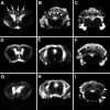
Figure 2. Brain MRI of Tgbov XV Mice Infected with BSE and BASE
T2-weighted images of anterior-to-posterior coronal planes of (A–C) BASE-infected mouse; (D–F) BSE-infected mouse; (G–I) uninfected Tgbov XV mouse. Both BSE- and BASE-infected mice show high signal intensity in the septal region: arrowheads in (A) and (D); and cerebellum: arrowheads in (C) and (F) compared to control (G) and (I). In addition, mice challenged with BASE exhibit scattered hyperintense areas in frontal regions: arrows in (A); and midbrain: arrows in (B) that are absent in BSE-infected and uninfected mice.
Figure 3
Figure 3. Survival Curves and PrPSc Types for Inbred Mice Following Primary Transmission of BSE and BASE
(A–D) Survival curves are for (A) SJL mice, (B) C57Bl/6 mice, (C) RIII mice, and (D) VM mice. (E) Western blot analysis of proteinase K-treated brain homogenates from SJL, RIII, C57Bl/6, and VM mice challenged with BSE. (F) Western blot analysis of brain homogenates from SJL, RIII, C57Bl/6, and VM mice challenged with BASE and a BSE-infected SJL mouse used as positive control (last lane). The first two lanes correspond to the same SJL mouse prior to and after proteinase K digestion, while all other samples were digested with proteinase K. The blots were probed with the anti-PrP antibody 6H4.
Figure 4
Figure 4. Lesion Profiles for Inbred Mice Following Primary Transmission of BSE
(A–D) Lesion profiles are for (A) SJL mice, (B) C57Bl/6 mice, (C) RIII mice, and (D) VM mice. The lesion profile was determined as specified in Figure 1. Data are mean ± s.e.m.
Figure 5
Figure 5. Survival Curves, Lesion Profiles, and PrPSc Types for Inbred Mice Following Second-Passage Transmission of BSE and BASE
(A and D) Survival curves are for (A) SJL mice and (D) C57Bl/6 mice. (B and E) Lesion profiles are for (B) SJL mice and (E) C57Bl/6 mice. The lesion profile was determined as specified in Figure 1. Data are mean ± s.e.m. (C and F) Western blot analysis of proteinase K-treated brain homogenates from SJL mice (C) and C57Bl/6 mice (F) after challenge with BSE and BASE. The blots were probed with the anti-PrP antibody 6H4.
Figure 6
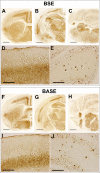
Figure 6. Immunohistochemical Detection of PrP in Brains of C57Bl/6 Mice Following Second-Passage Transmission of BSE and BASE
(A–E) Mice infected with mouse-derived BSE or (F–J) mouse-derived BASE show the same brain regional distribution and patterns of deposition of PrPSc. Micrographs were obtained from anterior-to-posterior coronal sections of the brain (A–C and F–H) and from corresponding areas of cerebral cortex (D and I) and cerebellum (E and J) probed with the anti-PrP antibody 6H4. Scale bar: 1 mm (A–C and F–H); 200 μm (D, E, I, and J).

References

    1. Prusiner SB. Prions. Proc Natl Acad Sci U S A. 1998;95:13363–13383. - PMC - PubMed
    1. Prusiner SB. Novel proteinaceous infectious particles cause scrapie. Science. 1982;216:136–144. - PubMed
    1. Kocisko DA, Come JH, Priola SA, Chesebro B, Raymond GJ, et al. Cell-free formation of protease-resistant prion protein. Nature. 1994;370:419–420. - PubMed
    1. Castilla J, Saa P, Hetz C, Soto C. In vitro generation of infectious scrapie prions. Cell. 2005;121:195–206. - PubMed
    1. Dickinson AG, Meikle VM. Host-genotype and agent effects in scrapie incubation: Change in allelic interaction with different strains of agent. Mol Gen Genet. 1971;112:73–79. - PubMed

Publication types

MeSH terms