. 2007;8(3):R37.
doi: 10.1186/gb-2007-8-3-r37.

Repetitive DNA is associated with centromeric domains in Trypanosoma brucei but not Trypanosoma cruzi

Affiliations

Repetitive DNA is associated with centromeric domains in Trypanosoma brucei but not Trypanosoma cruzi

Samson O Obado et al. Genome Biol. 2007.

Abstract

Background: Trypanosomes are parasitic protozoa that diverged early from the main eukaryotic lineage. Their genomes display several unusual characteristics and, despite completion of the trypanosome genome projects, the location of centromeric DNA has not been identified.

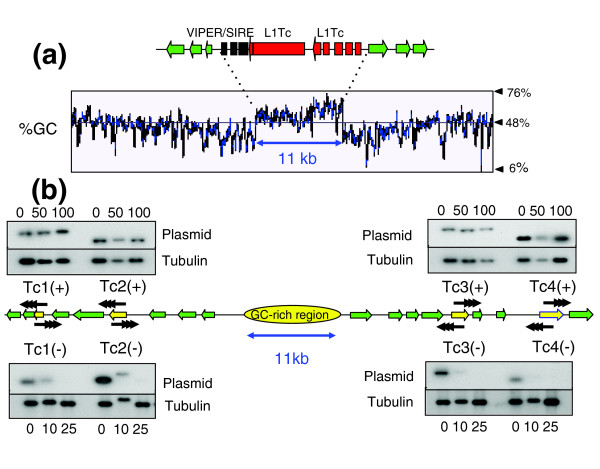
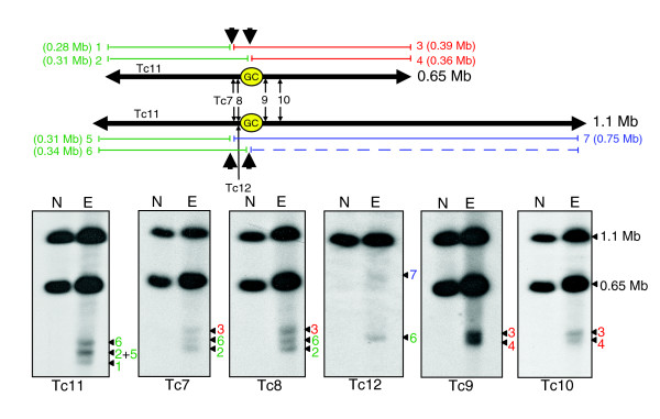
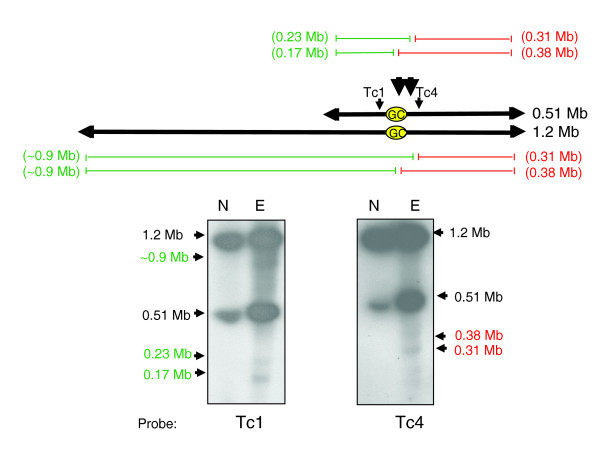
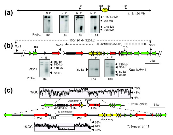
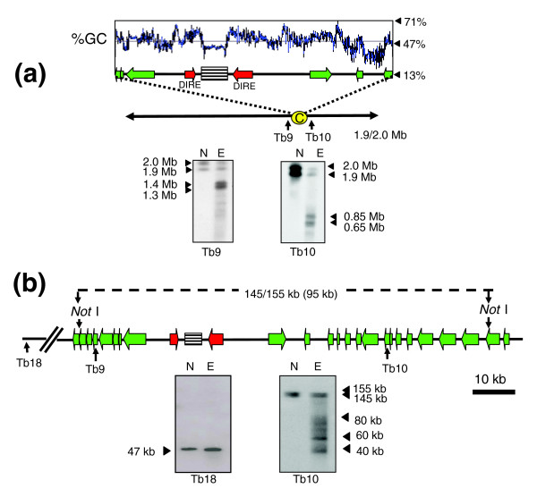
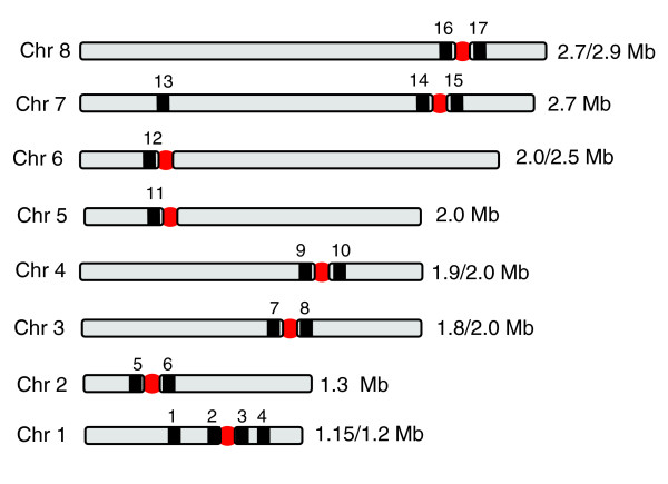
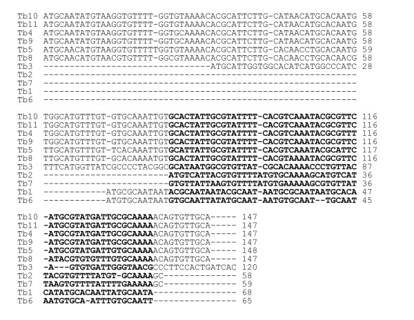
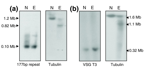
Results: We report evidence on the location and nature of centromeric DNA in Trypanosoma cruzi and Trypanosoma brucei. In T. cruzi, we used telomere-associated chromosome fragmentation and found that GC-rich transcriptional 'strand-switch' domains composed predominantly of degenerate retrotranposons are a shared feature of regions that confer mitotic stability. Consistent with this, etoposide-mediated topoisomerase-II cleavage, a biochemical marker for active centromeres, is concentrated at these domains. In the 'megabase-sized' chromosomes of T. brucei, topoisomerase-II activity is also focused at single loci that encompass regions between directional gene clusters that contain transposable elements. Unlike T. cruzi, however, these loci also contain arrays of AT-rich repeats stretching over several kilobases. The sites of topoisomerase-II activity on T. brucei chromosome 1 and T. cruzi chromosome 3 are syntenic, suggesting that centromere location has been conserved for more than 200 million years. The T. brucei intermediate and minichromosomes, which lack housekeeping genes, do not exhibit site-specific accumulation of topoisomerase-II, suggesting that segregation of these atypical chromosomes might involve a centromere-independent mechanism.

Conclusion: The localization of centromeric DNA in trypanosomes fills a major gap in our understanding of genome organization in these important human pathogens. These data are a significant step towards identifying and functionally characterizing other determinants of centromere function and provide a framework for dissecting the mechanisms of chromosome segregation.


Figures

Figure 1
Figure 1
Functional mapping of the putative centromere on T. cruzi chromosome 1. (a) Organization of the GC-rich strand-switch region. Green arrows identify ORFs in the polycistronic gene clusters and the implied direction of transcription. The degenerate retrotransposon-like VIPER/SIRE element (black) and L1Tc autonomous retroelements (red) are indicated. The %GC content was determined by the Artemis 7 program [38]. (b) Mitotic stability of truncated chromosomes. Sequences used for fragmentation (Tc1-Tc4) are indicated by yellow arrows. Vectors were targeted in both directions (+/-), with black arrowheads representing the positions and orientations of de novo telomeres after fragmentation (see Additional data files 1-3 for further details). Clones with truncated chromosomes were grown in the absence of G418 for the generations indicated above or below the corresponding track. Genomic DNA was ScaI digested, Southern blotted, probed with plasmid DNA, then re-hybridized with β-tubulin as a loading control.
Figure 2
Figure 2
Etoposide-mediated cleavage sites in T. cruzi chromosome 3. Epimastigotes were treated with 1 mM etoposide for 6 h and chromosomal DNA fractionated by CHEFE and assessed by Southern analysis. Probe Tc12 is specific to the larger homologue (see Materials and methods). Lane N, non-treated parasites; lane E, etoposide-treated. The schematic shows both chromosome 3 homologues, location of the 16 kb GC-rich strand-switch domain (GC), positions of the probes and predicted locations of the major Topo-II cleavage sites (large black arrowheads). The fragments generated (1-7), and their sizes and inferred positions on the chromosomes are shown in red, green and blue. With the exception of probe Tc12, fragments derived from the right arm of the 1.1 Mb homologue (blue) cannot be detected, due to co-migration with the cross-hybridizing 0.65 Mb homologue.
Figure 3
Figure 3
Mapping of etoposide-mediated Topo-II cleavage sites in T. cruzi chromosome 1. Epimastigotes were treated with 1 mM etoposide for 6 h and chromosomal DNA fractionated by CHEFE and assessed by Southern hybridization. Probes Tc1 and Tc4 were used (Additional data file 5). Large black arrowheads identify the predicted locations of Topo-II activity adjacent to the GC-rich strand-switch domain (yellow oval). The cleavage fragments are identified in red and green. Lane N, non-treated parasites; lane E, etoposide-treated.
Figure 4
Figure 4
Etoposide-mediated cleavage sites on T. brucei chromosome 1. (a) Procyclics were treated with 500 μM etoposide for 1 h and chromosomal DNA fractionated by CHEFE. Hybridization was carried out with probes Tb1-Tb4. Their positions and the location of the strand-switch domain (yellow oval) are shown. Lane N, non-treated parasites; lane E, etoposide-treated. (b) Fine-mapping of cleavage sites. Chromosomal DNA from treated/non-treated parasites was immobilized in agar blocks, restriction digested and fractionated by CHEFE. Fragment sizes are shown above the schematic, with their predicted sizes (GeneDB) in parentheses. Black triangles identify the fragments and cleavage products on the relevant autoradiographs. As control, blots were re-hybridized with probe Tb4, from a gene 150 kb upstream of the putative centromere. (c) Comparison of the T. cruzi chromosome 3 centromeric domain with the syntenic region of T. brucei chromosome 1. In the T. cruzi chromosome, the degenerate VIPER/SIRE element and L1Tc retroelements are indicated, together with a truncated cruzipain pseudogene (ψCZP) and an U2snRNA gene. The corresponding region in T. brucei chromosome 1 contains 2 INGI retrotransposons and 1 DIRE. The locations of a leucine rich repeat protein gene (LRRP), a rRNA gene array and a 5.5 kb array of approximately 30 bp repeats are shown. Green arrows indicate putative ORFs and the implied direction of transcription. The dashed lines between the T. cruzi and T. brucei maps identify the equivalent positions of the first ORFs of the conserved directional gene clusters.
Figure 5
Figure 5
Etoposide-mediated cleavage sites on T. brucei chromosome 4. (a) Procyclics were treated with 500 μM etoposide for 1 h and chromosomal DNA fractionated by CHEFE and Southern blotted. Red arrows identify DIREs and green arrows indicate putative ORFs and the implied direction of transcription. The location of the AT-rich repeat array is highlighted (striped box). The positions of probes and location of the putative centromeric region (yellow oval) are indicated. Lane N, non-treated parasites; lane E, etoposide-treated. (b) Fine mapping of cleavage sites. NotI digested DNA was fractionated by CHEFE as in Figure 4b and Southern blotted. Fragment sizes are shown above the schematic, with their predicted sizes (GeneDB) in parentheses. Black triangles identify these fragments on the autoradiograph and show the major cleavage products. As control, the membrane was hybridized with probe Tb18, from a gene 500 kb downstream of the putative centromere.
Figure 6
Figure 6
Etoposide-mediated Topo-II cleavage sites (red circles) on T. brucei (strain 927) chromosomes 1-8. The locations of the probes (Tb1-17) are identified by black bars. Details on the AT-rich arrays are given in Figure 7 and Table 1 and the experimental results are shown in Additional data file 4.
Figure 7
Figure 7
Alignment of repeat array consensus sequences on each T. brucei chromosome, determined by the program Tandem Repeats Finder [39]. The nucleotides marked in bold correspond to two copies of the array (approximately 30 bp) in chromosomes 1, 2, 6 and 7.
Figure 8
Figure 8
T. brucei (a) minichromosomes and (b) intermediate chromosomes are resistant to etoposide-mediated cleavage. (a) Procyclics (927 strain) were treated with 500 μM etoposide for 3 h and chromosomal DNA separated by CHEFE. Southern blots were hybridized with the 177 bp repeat probe or α-tubulin. (b) Bloodstream form T. brucei (427 strain) were treated with 25 μM etoposide for 1 h and chromosomal DNA analyzed as above. Blots were hybridized with the intermediate chromosome-specific probe T3. In strain 427, chromosome 1 is larger than in strain 927, as is the major cleavage product identified with the α-tubulin probe. Lane N, non-treated parasites; lane E, etoposide-treated.

References

    1. Fukagawa T. Centromere DNA, proteins and kinetochore assembly in vertebrate cells. Chromosome Res. 2004;12:557–567. doi: 10.1023/B:CHRO.0000036590.96208.83. - DOI - PubMed
    1. Steiner NC, Hahnenberger KM, Clarke L. Centromeres of the fission yeast Schizosaccharomyces pombe are highly variable genetic loci. Mol Cell Biol. 1993;13:4578–4587. - PMC - PubMed
    1. Pidoux AL, Allshire RC. Kinetochore and heterochromatin domains of the fission yeast centromere. Chromosome Res. 2004;12:521–534. doi: 10.1023/B:CHRO.0000036586.81775.8b. - DOI - PubMed
    1. Maddox PS, Oegema K, Desai A, Cheeseman IM. 'Holo'er than thou: chromosome segregation and kinetochore function in C. elegans. Chromosome Res. 2004;12:641–653. doi: 10.1023/B:CHRO.0000036588.42225.2f. - DOI - PubMed
    1. Schueler MG, Higgins AW, Rudd MK, Gustashaw K, Willard HF. Genomic and genetic definition of a functional human centromere. Science. 2001;294:109–115. doi: 10.1126/science.1065042. - DOI - PubMed

Publication types

LinkOut - more resources