. 2007 May;27(10):3881-90.
doi: 10.1128/MCB.02366-06. Epub 2007 Mar 12.

trans Autophosphorylation at DNA-dependent protein kinase's two major autophosphorylation site clusters facilitates end processing but not end joining

Affiliations

trans Autophosphorylation at DNA-dependent protein kinase's two major autophosphorylation site clusters facilitates end processing but not end joining

Katheryn Meek et al. Mol Cell Biol. 2007 May.

Abstract

Recent studies have established that DNA-dependent protein kinase (DNA-PK) undergoes a series of autophosphorylation events that facilitate successful completion of nonhomologous DNA end joining. Autophosphorylation at sites in two distinct clusters regulates DNA end access to DNA end-processing factors and to other DNA repair pathways. Autophosphorylation within the kinase's activation loop regulates kinase activity. Additional autophosphorylation events (as yet undefined) occur that mediate kinase dissociation. Here we provide the first evidence that autophosphorylation within the two major clusters (regulating end access) occurs in trans. Further, both UV-induced and double-strand break (DSB)-induced phosphorylation in the two major clusters is predominantly autophosphorylation. Finally, we show that while autophosphorylation in trans on one of two synapsed DNA-PK complexes facilitates appropriate end processing, this is not sufficient to promote efficient end joining. This suggests that end joining in living cells requires additional phosphorylation events that either occur in cis or that occur on both sides of the DNA-PK synapse. These data support an emerging consensus that, via a series of autophosphorylation events, DNA-PK undergoes a sequence of conformational changes that promote efficient and appropriate repair of DSBs.


Figures

FIG. 1.
FIG. 1.
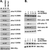
DNA DSBs and DNA cross-links induce ABCDE and PQR autophosphorylation in living cells. A. Diagrammatic representation of DNA-PKcs. Autophosphorylation sites are denoted with asterisks. Thirteen previously described autophosphorylation sites [A, B, C, D, E, M, P, Q, R, and T] are as follows: A = T2609, B = T2620 + S2624, C = T2638, D = T2647, E = S2612, M = S3205, P = S2023 + S2029; Q = S2041, R = S2056 + S2053, and T = T3950. LRR denotes the leucine-rich region (13). The position of splice variation (7) in the phosphatidylinositol 3-kinase (PI3K) domain is shown. Isoform II lacks residues 3883 to 4128; isoform III lacks residues 3797 to 3828. Conserved FAT and FAT-C domains denote regions in the C terminus conserved in ATM, ATR, DNA-PKcs, and related kinases. ATP denotes a lysine residue (K3752) adjacent to the proposed ATP binding site. B. Cell extracts (100 μg) from V3 transfectants expressing wild-type DNA-PKcs (lane 1), ABCDE mutant DNA-PKcs (lane 2), or PQR mutant DNA-PKcs (lane 3) were incubated under kinase-active conditions for 20 min at room temperature. Kinase reactions were then analyzed by immunoblotting with a phospho-specific antibody that recognizes phospho-A (2609, top panel), phospho-R (2056, middle panel), or total DNA-PKcs (bottom panel). C. V3 transfectants expressing wild-type DNA-PKcs were not treated (lanes 1 and 4) or treated with OA (lanes 2 and 5), zeocin (Zeo) and OA (lane 3), MMC (lane 6), or MMC and OA (lane 7) as indicated. Cells were harvested 1 hour later, and cell extracts were analyzed as described above.
FIG. 2.
FIG. 2.
DNA-PKcs primarily autophosphorylates A (2609) and R (2056) in response to DSBs. A. Stable V3 transfectants expressing wild-type DNA-PKcs (lanes 1 and 2), ABCDE+PQR mutant DNA-PKcs (lanes 3 and 4), or K>R mutant DNA-PKcs (lanes 5 and 6) were treated or not with 30 J UV and OA. After 1 h, cell extracts were prepared and analyzed by immunoblotting as in Fig. 1B. This is representative of five independent experiments. In UV experiments, low levels of A (2609) and R (2056) phosphorylation were detected in untreated cells and were presumably an artifact that resulted from mock UV treatment of cells in the absence of medium or PBS. B. Stable V3 transfectants expressing wild-type DNA-PKcs (lanes 1 and 2) or K>R mutant DNA-PKcs (lanes 3 and 4) were treated or not with zeocin (Z) and OA. After 1 h, cell extracts were prepared and analyzed by immunoblotting as described for Fig. 1B.
FIG. 3.
FIG. 3.
In human cells, DNA-PKcs primarily autophosphorylates the ABCDE, PQR, and T sites in response to DSBs. A. Immunoblotting of DNA-PKcs partially purified from HEK293 cells treated or not with IR and with the ATM-specific inhibitor and the DNA-PKcs specific inhibitor as described in Materials and Methods. Phospho-specific antibodies recognized phosphorylated R (2056), A (2609), E (2612), B (2620 and 2624), C (2638), D (2647), and T (3950). B. Immunoblotting with ATM, p53, Chk2, actin, and DNA-PKcs from HEK293 cells treated or not with IR and with the ATM-specific inhibitor KU55933 as described in Materials and Methods. Phospho-specific antibodies recognized phosphorylated S1981 in ATM, phosphorylated S15 in p53, phosphorylated T68 in Chk2, and phosphorylated R (2056) in DNA-PKcs.
FIG. 4.
FIG. 4.
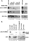
ABCDE and PQR autophosphorylation can occur in trans in vitro and in living cells. A. WCE from V3 transfectants expressing wild-type DNA-PKcs (lane 1), the ABCDE+PQR mutant (lane 2), the ATP binding site mutant K>R (lane 4), or both (lane 3) were incubated in the presence of ATP and calf thymus DNA as described in Materials and Methods. Phosphorylation at the R site (2056) or the A site (2609) was detected with phospho-specific antibodies, as indicated. B. Cells expressing wild-type DNA-PKcs (lanes 1 and 2), GFP-K>R (lanes 3 and 4), or the ABCDE+PQR combined mutant (lanes 5 to 10) were treated or not with zeocin (Zeo). The ABCDE+PQR mutant was either untransfected (lanes 5 and 6) or transiently transfected with plasmids encoding the GFP-K>R mutant DNA-PKcs (lanes 7 and 8) or GFP-tagged wild-type DNA-PKcs (lanes 9 and 10). After a 1-hour incubation with zeocin, cell extracts were analyzed by immunoblotting with the 2056 phospho-specific antibody (top panel) or a GFP-specific antibody (bottom panel). (Note: signal in untagged wild-type DNA-PKcs [lane 2, bottom panel] is residual phospho-R reactivity from the initial probing.)
FIG. 5.
FIG. 5.
The C terminus of DNA-PKcs may be important for DNA-PK synapsis. A. WCE from V3 transfectants expressing wild-type DNA-PKcs (lanes 1 and 11), the ABCDE+PQR mutant (lanes 2 and 12), the ATP binding site mutant K>R (lane 7), kinase-inactive isoform III (lane 8), kinase-inactive isoform II (lane 9), the kinase-impaired 3′ mutant (lane 10; this mutation is representative of the DNA-PKcs mutation in the DSBR mutant cell strain XR-C2), kinase-inactive mutant T>D (lane 14), or combinations of the ABCDE+PQR mutant with kinase-inactive mutants (lanes 3 to 6 and 13) as indicated were incubated in the presence of ATP and calf thymus DNA as described in Materials and Methods. Phosphorylation at the R site (2056) or the A site (2609) was detected with phospho-specific antibodies as indicated. B. The ABCDE+PQR mutant was either untransfected (lanes 3 and 4) or transiently transfected with plasmids encoding wild-type GFP-DNA-PKcs (lanes 1 and 2), GFP-K>R mutant DNA-PKcs (lanes 5 and 6), GFP-isoform III (lanes 7 and 8), untagged T>D mutant (lanes 9 and 10), FLAG-isoform II (lanes 11 and 12), or FLAG-wild-type DNA-PKcs (lanes 13 and 14). Cells were treated or not with zeocin (Z) as indicated. After 1 hour, cells were harvested and extracts analyzed by immunoblotting with a GFP-specific antibody (top panel) or the 2056 phospho-specific antibody (bottom panel).
FIG. 6.
FIG. 6.
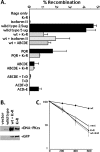
trans autophosphorylation of ABCDE or PQR on one of two synapsed DNA-PK complexes cannot rescue the end-joining deficits associated with autophosphorylation blockade or kinase activity blockade. RAG expression from plasmid vectors initiates recombination in the V3 cells, as assessed by the plasmid substrate pJH290, which detects coding joints. DNA-PKcs-encoding plasmids were included in the transient assay as indicated (described in Materials and Methods). Recombination rates from four to nine independent assays were averaged. Error bars denote standard deviations.
FIG. 7.
FIG. 7.
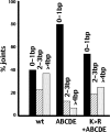
Nucleotide nibbling is not asymmetric in joints mediated by the ABCDE+K>R synapse. Sequences from wild-type-only, ABCDE-only, or ABCDE+K>R transfections (summarized in Table 1; data available on request) were analyzed as follows. All sequences that have one end with 3 bp or more deleted were grouped, and the number of the base pair missing from the second end was tabulated and grouped as follows: 0 to 1 bp (black bars); 2 to 3 bp (striped bars); 4 bp or more (stippled bars). Numbers presented are the percentages of joints with 3 bp or more deleted from one end.

Similar articles

Cited by

References

    1. Block, W., Y. Yu, D. Merkle, J. Gifford, Q. Ding, K. Meek, and S. P. Lees-Miller. 2004. Autophosphorylation-dependent remodeling of the DNA-dependent protein kinase catalytic subunit (DNA-PKcs) regulates ligation of DNA ends. Nucleic Acids Res. 32:4351-4357. - PMC - PubMed
    1. Boskovic, J., A. Rivera-Calzada, J. D. Maman, P. Chacon, K. R. Willison, L. H. Pearl, and O. Llorca. 2003. Visualization of DNA-induced conformational changes in the DNA repair kinase DNA-PKcs. EMBO J. 22:5875-5882. - PMC - PubMed
    1. Chan, D. W., B. P. Chen, S. Prithivirajsingh, A. Kurimasa, M. D. Story, J. Qin, and D. J. Chen. 2002. Autophosphorylation of the DNA-dependent protein kinase catalytic subunit is required for rejoining of DNA double-strand breaks. Genes Dev. 16:2333-2338. - PMC - PubMed
    1. Chan, D. W., and S. P. Lees-Miller. 1996. The DNA-dependent protein kinase is inactivated by autophosphorylation of the catalytic subunit. J. Biol. Chem. 271:8936-8941. - PubMed
    1. Chen, B. P., N. Uematsu, J. Kobayashi, Y. Lerenthal, A. Krempler, H. Yajima, M. Lobrich, Y. Shiloh, and D. J. Chen. 2006. Ataxia telangiectasia mutated (ATM) is essential for DNA-PKcs phosphorylations at the Thr-2609 cluster upon DNA double strand break. J. Biol. Chem. 282:6582-6587. - PubMed

Publication types