. 2007 May;27(10):3708-15.
doi: 10.1128/MCB.01838-06. Epub 2007 Mar 12.

A novel Stat3 binding motif in Gab2 mediates transformation of primary hematopoietic cells by the Stk/Ron receptor tyrosine kinase in response to Friend virus infection

Affiliations

A novel Stat3 binding motif in Gab2 mediates transformation of primary hematopoietic cells by the Stk/Ron receptor tyrosine kinase in response to Friend virus infection

Shuang Ni et al. Mol Cell Biol. 2007 May.

Abstract

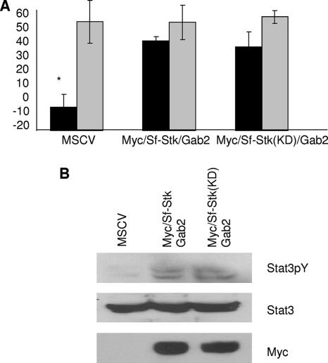
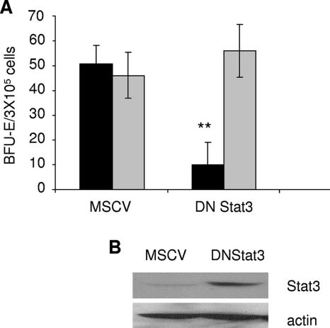
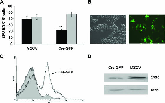
Friend erythroleukemia virus has long served as a paradigm for the study of the multistage progression of leukemia. Friend virus infects erythroid progenitor cells, followed by an initial polyclonal expansion of infected cells, which is driven by the activation of a naturally occurring truncated form of the Stk receptor tyrosine kinase (Sf-Stk). Subsequently, the accumulation of additional mutations in p53 and the activation of PU.1 result in full leukemic transformation. The early stages of transformation induced by Friend virus are characterized in vitro by the Epo-independent growth of infected erythroblasts. We have shown previously that this transforming event requires the kinase activity and Grb2 binding site of Sf-Stk and the recruitment of a Grb2/Gab2 complex to Sf-Stk. Here, we demonstrate that Stat3 is required for the Epo-independent growth of Friend virus-infected cells and that the activation of Stat3 by Sf-Stk is mediated by a novel Stat3 binding site in Gab2. These results underscore a central role for Stat3 in hematopoietic transformation and describe a previously unidentified role for Gab2 in the recruitment and activation of Stat3 in response to transforming signals generated by tyrosine kinases.


Figures

FIG. 1.
FIG. 1.
The region between Q120 and P358 of Gab2 is essential to support the transformation of Friend virus-infected erythroblasts. (A) Schematic diagram of the chimeric Gab1/2 constructs used in this study. Gray bars, sequence from Gab1; black bars, sequence from Gab2. (B) Total bone marrow from Gab2−/− mice was transduced with empty vector (MSCV), Gab2, and the indicated Gab1/2 chimeras (CM1 to CM6). Transduced cells were plated in methylcellulose containing IL-3 (2.5 ng/ml) in the presence of FV or Epo (1 U/ml). BFU-E cells were stained with acid-benzidine and scored on day 5. Black columns, Friend virus; gray columns, Epo. Bars denote the means ± SD of one of three independent experiments performed in replicates of three and reflect normalized values. *, P < 0.05; **, P < 0.01. (C) Expression of Gab1, Gab2, and Gab1/2 chimeric proteins in 293T cells, determined by Western blot analysis using a mixture of anti-Gab1 and anti-Gab2 antibodies.
FIG. 2.
FIG. 2.
The region between Q120 and P358 of Gab2 is required to mediate Stat3 activation when coexpressed with Sf-Stk. 293T cells were transiently cotransfected with Stat3 and plasmids expressing (A) wild-type or mutant forms of Sf-Stk or (B) wild-type or mutant forms of Sf-Stk/Gab fusions as indicated. Cell lysates were probed with anti-phospho-Stat3, anti-Stat3, and anti-Myc. (C) 293T cells were transiently cotransfected with Myc-Sf-Stk, Stat3, and plasmids expressing the various Gab1/2 fusions as indicated. Cell lysates were probed with anti-phospho-Stat3, anti-Stat3, and anti-Gab1/2.
FIG. 3.
FIG. 3.
The ability of Gab2 to recruit and activate Stat3 is dependent on the Y194LHQ site in Gab2. (A) Sequence alignment of mGab1 and mGab2 proteins. The potential Stat3 binding site is shown in the box. (B) 293T cells were transiently cotransfected with Stat3 and plasmids expressing wild-type (MSCV) or mutant Sf-Stk/Gab2 fusions as indicated. Cell lysates were immunoprecipitated (IP) with anti-Stat3 antibodies and subsequently analyzed by SDS-PAGE and Western immnunoblotting (IB) with anti-Myc and anti-Stat3 antibodies. (C) 293T cells were transiently cotransfected with Stat3, Sf-Stk, and wild-type or mutant forms of Gab2. Cell lysates were immunoprecipitated with anti-Stat3 and blotted with anti-Gab2, anti-Myc, and anti-Stat3 antibodies. −, absence of; +, presence of.
FIG. 4.
FIG. 4.
The Y194LHQ site in Gab2 is critical to support Epo-independent colony formation of primary erythroblasts in response to Friend virus infection. (A) Total bone marrow from C57BL/6 mice was transduced with empty vector (MSCV), Myc-Sf-Stk/Gab2, or Myc-Sf-Stk/Gab2 harboring the Y194-to-F mutations. Transduced cells were plated in methylcellulose containing IL-3 (2.5 ng/ml) in the presence or absence of FV or Epo (1 U/ml). BFU-E cells were stained with acid-benzidine and scored on day 5. Black columns, Friend virus; gray columns, Epo. Bars denote the means ± SD of one of three independent experiments performed in replicates of three and reflect normalized values. (B) Expression of Myc-sf-stk/Gab2 and Myc-sf-stk/Gab2(Y/F) in 293T packaging cells. (C) Total bone marrow from Gab2−/− mice was transduced with empty vector, Gab2 or Gab2(Y194/F). Colony assays were performed as described in the legend for panel A. (D) Expression of Gab2 and Gab2(Y/F) in 293T packaging cells. *, P < 0.05; **, P < 0.01.
FIG. 5.
FIG. 5.
The kinase activity of Sf-Stk is not required for the phosphorylation of Stat3 or Epo-independent colony formation of primary erythroblasts in response to Friend virus infection. (A) Total bone marrow from Gab2−/− C57BL/6 mice was transduced with empty vector (MSCV), Myc-Sf-Stk/Gab2, or Myc-Sf-Stk(KD)/Gab2. Transduced cells were plated in methylcellulose containing IL-3 (2.5 ng/ml) in the presence or absence of FV or Epo (1 U/ml). BFU-E cells were stained with acid-benzidine and scored on day 5. Black columns, Friend virus; gray columns, Epo. Bars denote the means ± SD of one of three independent experiments performed in replicates of three and reflect normalized values. *, P < 0.05. (B) 293T cells were transiently cotransfected with Stat3 and plasmids indicated above. Cell lysates were probed with anti-phospho-Stat3, anti-Stat3, and anti-Myc.
FIG. 6.
FIG. 6.
Interaction of Gab2 with Stat3 and tyrosine phosphorylation of Stat3 in spleens of Friend virus-infected mice. (A) Ten-week-old BALB/cJ mice were injected in their tail veins with FVP at different time points (day 14, day 10, day 7, and day 4). Spleen protein was harvested and subsequently analyzed by SDS-PAGE and Western blotting with anti-phospho-Stat3(Y705), anti-Stat3, and anti-phospho-Stat3(S727). (B) Ten-week-old BALB/cJ mice were injected in their tail veins with FVP at different time points (day 10 and day 7). Spleen protein was immunoprecipitated with anti-Gab2 antibodies and subsequently analyzed by SDS-PAGE and Western blotting with anti-Stat3 and anti-Gab2.
FIG. 7.
FIG. 7.
Expression of dominant negative Stat3 inhibits colony formation in response to Friend virus in vitro. (A) Total bone marrow from BALB/c mice was transduced with empty vector (MSCV) or a dominant negative Stat3 [DNStat3(Y705F)]. Transduced cells were plated in methylcellulose containing IL-3 (2.5 ng/ml) in the presence of FV or Epo (1 U/ml). BFU-E cells were stained with acid-benzidine and scored on day 5. Black columns, Friend virus; gray columns, Epo. Bars denote the means ± SD of one of three independent experiments performed in replicates of three and reflect normalized values. **, P < 0.01. (B) Expression of dominant negative Stat3 in 293T packaging cells.
FIG. 8.
FIG. 8.
Expression of Cre recombinase inhibits colony formation by bone marrow cells from floxed Stat3 mice in response to Friend virus infection. (A) Total bone marrow from floxed Stat3 BALB/c mice was transduced with empty vector or a Cre-GFP fusion plasmid. Transduced cells were plated in methylcellulose containing IL-3 (2.5 ng/ml) in the presence of FV or Epo (1 U/ml). BFU-E cells were stained with acid-benzidine and scored on day 5. Black columns, Friend virus; gray columns, Epo. Bars denote the means ± SD of one of three independent experiments performed in replicates of three and reflect normalized values. **, P < 0.01. (B) Expression of Cre-GFP in 293T packaging cells by immunofluorescence. (C) Total bone marrow from floxed Stat3 mice was infected with vector (shaded area) or Cre-GFP (solid line). Infection efficiency was determined by flow cytometry for GFP. (D) Expression of Stat3 in total bone marrow from floxed Stat3 mice infected with empty vector or Cre-GFP.

Similar articles

Cited by

References

    1. Bentires-Alj, M., S. G. Gil, R. Chan, Z. C. Wang, Y. Wang, N. Imanaka, L. N. Harris, A. Richardson, B. G. Neel, and H. Gu. 2006. A role for the scaffolding adapter GAB2 in breast cancer. Nat. Med. 12:114. - PubMed
    1. Besser, D., J. Bromberg, J. Darnell, and H. Hanafusa. 1999. A single amino acid substitution in the v-Eyk intracellular domain results in activation of Stat3 and enhances cellular transformation. Mol. Cell. Biol. 19:1401-1409. - PMC - PubMed
    1. Bromberg, J., C. Horvath, D. Besser, W. Latham, and J. Darnell. 1998. Stat3 activation is required for cellular transformation by v-src. Mol. Cell. Biol. 18:2553-2558. - PMC - PubMed
    1. Bromberg, J., M. Wrzeszczynska, G. Devgan, Y. Zhao, R. Pestell, C. Albanese, and J. Darnell. 1999. Stat3 as an oncogene. Cell 98:295-303. - PubMed
    1. Casteran, N., P. De Sepulveda, N. Beslu, M. Aoubala, S. Letard, E. Lecocq, R. Rottapel, and P. Dubreuil. 2003. Signal transduction by several KIT juxtamembrane domain mutations. Oncogene 22:4710-4722. - PubMed

Publication types

MeSH terms