. 2007 Jun;75(6):2668-78.
doi: 10.1128/IAI.01872-06. Epub 2007 Mar 12.

A genetic screen for Mycobacterium tuberculosis mutants defective for phagosome maturation arrest identifies components of the ESX-1 secretion system

Affiliations

A genetic screen for Mycobacterium tuberculosis mutants defective for phagosome maturation arrest identifies components of the ESX-1 secretion system

Jason A MacGurn et al. Infect Immun. 2007 Jun.

Abstract

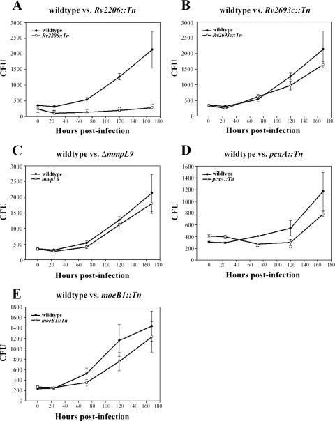
After phagocytosis, the intracellular pathogen Mycobacterium tuberculosis arrests the progression of the nascent phagosome into a phagolysosome, allowing for replication in a compartment that resembles early endosomes. To better understand the molecular mechanisms that govern phagosome maturation arrest, we performed a visual screen on a set of M. tuberculosis mutants specifically attenuated for growth in mice to identify strains that failed to arrest phagosome maturation and trafficked to late phagosomal compartments. We identified 10 such mutants that could be partitioned into two classes based on the kinetics of trafficking. Importantly, four of these mutants harbor mutations in genes that encode components of the ESX-1 secretion system, a pathway critical for M. tuberculosis virulence. Although ESX-1 is required, the known ESX-1 secreted proteins are dispensable for phagosome maturation arrest, suggesting that a novel effector required for phagosome maturation arrest is secreted by ESX-1. Other mutants identified in this screen had mutations in genes involved in lipid synthesis and secretion and in molybdopterin biosynthesis, as well as in genes with unknown functions. Most of these trafficking mutants exhibited a corresponding growth defect during macrophage infection, but two mutants grew like wild-type M. tuberculosis during macrophage infection. Our results support the emerging consensus that multiple factors from M. tuberculosis, including the ESX-1 secretion system, are involved in modulating trafficking within the host.


Figures

FIG. 1.
FIG. 1.
Identification of trafficking mutants in M. tuberculosis. Bone-marrow derived macrophages were infected with strains of M. tuberculosis and, at 24 and 72 h postinfection, pulsed for 10 min with fluorescent dextran immediately prior to fixation. Fixed monolayers were imaged by using fluorescence deconvolution microscopy. Fluorescent dextran is green, while bacilli were stained red. White arrows point to individual bacilli. (A) Live and heat-killed M. tuberculosis were used as controls. (B) The screen identified several mutants exhibiting aberrant dextran colocalization. (C and D) For each strain, the fraction colocalization at 24 and 72 h postinfection was averaged over at least three separate infections totaling >200 phagosomes. A total of 10 mutants with colocalization defects were identified. Scale bars, 10 μm. **, P < 0.001.
FIG. 2.
FIG. 2.
Colocalization of M. tuberculosis strains with different maturation markers. Infections were performed and imaged as in Fig. 1. Representative images for 24 h and 72 h postinfection are shown from macrophages infected with live and heat-killed M. tuberculosis, as well as Rv3877::Tn and Rv3615c::Tn mutant strains. Maturation markers are green, while bacilli were stained red. White arrows point to individual bacilli. (A) For fluorescent transferrin colocalization, infected monolayers were pulsed with fluorescent transferrin for 10 min immediately prior to fixation. (B and C) Fixed monolayers were stained for late compartment markers LBPA and Lamp-2 by indirect immunofluorescence. Scale bars, 10 μm.
FIG. 3.
FIG. 3.
In vivo growth defects of PMA mutant strains. (A) Array filters were used to determine tag representation within pools of signature-tagged transposon mutants from culture (input) and infected mice (output). WT, a strain in the pool of mutants that exhibited wild-type growth in mice at 3 weeks postinfection. (B) For each spot on the array filters, representation was determined by densitometry. For each mouse, the following equation was used to calculate a ratio of pixel intensities normalized to the wild type: (mutantmouse/wild typemouse)/(mutantinput/wild typeinput).
FIG. 4.
FIG. 4.
Analysis of intracellular survival of trafficking mutants. Bone marrow-derived macrophage monolayers were infected with M. tuberculosis strains at an MOI of 1 for 2 h and plated for CFU determination at 0, 24, 72, 120, and 168 h postinfection. CFU were determined for the Rv2206::Tn (A), Rv2693::Tn (B), ΔmmpL9 (C), pcaA::Tn (D), and moeB1::Tn (E) mutants. Values shown on the y axis are from a dilution of the monolayer lysate and represent 1/400 of the actual number of CFU in the monolayer. Growth curves shown are representative growth curves from at least two similar experiments. *, P < 0.05; **, P < 0.01.
FIG. 5.
FIG. 5.
Quantification of trafficking defects of ESX-1 mutants. Several mutants known to be defective for ESX-1 secretion (Rv3877::Tn, Rv3870::Tn, Rv3871::Tn, ΔesxA, and Rv3615c::Tn) were scored for their ability to mediate PMA at 24 h and 72 h postinfection. (A to D) A short dextran pulse (A) and transferrin (B) were used as markers of early phagosomal compartments, whereas LBPA (C) and Lamp-2 (D) were used as markers of late phagosomal compartments. For each strain, fraction colocalization was averaged over at least three separate infections totaling >200 phagosomes. **, significant difference (P < 0.01) compared to both wild-type and ΔesxA strains.
FIG. 6.
FIG. 6.
ΔesxA is not defective for PMA and does not traffic to a late phagosome. (A) Macrophages were infected with M. tuberculosis ΔesxA mutant and analyzed for colocalization with transferrin and LBPA by using fluorescence deconvolution microscopy. Fluorescent markers are green, while bacilli were stained red. White arrows point to individual bacilli. Scale bar, 10 μm. (B) differential interference contrast (left), deconvolved fluorescent z-slice (middle), and merged (right) images of individual macrophages infected with either Rv3877::Tn or ΔesxA mutant strains were stained for the late marker LBPA by indirect immunofluorescence. Images are from 24 h postinfection. Nuclei (blue) were stained with DAPI (4′,6′-diamidino-2-phenylindole). (C) Z-stacks from macrophages infected with either Rv3877::Tn or ΔesxA mutants (stained red) were deconvolved and rendered to visualize and determine colocalization in three dimensions. LBPA stain is green. (D) Rv3877::Tn and ΔesxA mutants (green) expressing green fluorescent protein were also used in some experiments as an alternative to postfixation staining of bacteria. The LBPA stain is red.

References

    1. Armstrong, J. A., and P. D. Hart. 1971. Response of cultured macrophages to Mycobacterium tuberculosis, with observations on fusion of lysosomes with phagosomes. J. Exp. Med. 134:713-740. - PMC - PubMed
    1. Behr, M. A., M. A. Wilson, W. P. Gill, H. Salamon, G. K. Schoolnik, S. Rane, and P. M. Small. 1999. Comparative genomics of BCG vaccines by whole-genome DNA microarray. Science 284:1520-1523. - PubMed
    1. Berthet, F. X., P. B. Rasmussen, I. Rosenkrands, P. Andersen, and B. Gicquel. 1998. A Mycobacterium tuberculosis operon encoding ESAT-6 and a novel low-molecular-mass culture filtrate protein (CFP-10). Microbiology 144(Pt. 11):3195-3203. - PubMed
    1. Chua, J., I. Vergne, S. Master, and V. Deretic. 2004. A tale of two lipids: Mycobacterium tuberculosis phagosome maturation arrest. Curr. Opin. Microbiol. 7:71-77. - PubMed
    1. Clemens, D. L., and M. A. Horwitz. 1995. Characterization of the Mycobacterium tuberculosis phagosome and evidence that phagosomal maturation is inhibited. J. Exp. Med. 181:257-270. - PMC - PubMed

Publication types

LinkOut - more resources