

Review
Neuropathology for the neuroradiologist: fluorescence in situ hybridization
Affiliations
- PMID: 17353304
- PMCID: PMC7977815
Item in Clipboard
Review
Neuropathology for the neuroradiologist: fluorescence in situ hybridization
AJNR Am J Neuroradiol.
2007 Mar.
Abstract
Fluorescence in situ hybridization is a molecular cytogenetic technique that localizes segments of DNA within tumor cells by using dyes that are visible with a fluorescent microscope. The technique has proved useful in typing a variety of tumors such as oligodendrogliomas and in understanding the genetic forces driving oncogenesis.
Figures
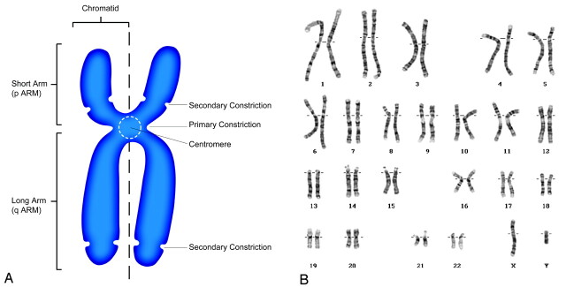
The human chromosome. A, Schematic drawing of a human chromosome in metaphase of the cell cycle. The chromosome consists of chromatin, which is made of a molecule of DNA complexly coiled around a protein frame, forming a chromatid. Paired chromatids, consisting of identical molecules of DNA, are joined at the centromere. B, Normal male karyotype showing the 23 pairs of human chromosomes for a total of 46 arranged in 8 groups based on size and shape. Karyotype courtesy of Colorado Genetic Laboratory, University of Colorado Health Sciences Center, Denver.
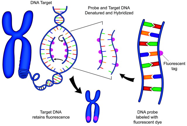
Schematic representation of FISH technique. A DNA probe is tagged with a fluorescent marker. The probe and target DNA are denatured, and the probe is allowed to hybridize with the target. The fluorescent tag is then detected with a fluorescent microscope.
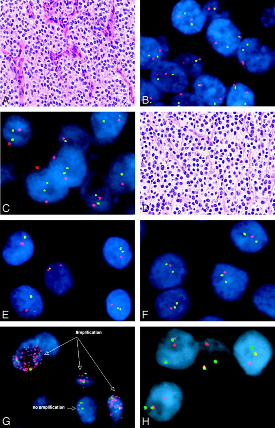
Representative FISH studies in tumors of the nervous system—central neurocytoma (A–C), oligodendroglioma (D–F), small cell glioblastoma (G), and Ewing sarcoma/peripheral primitive neuroectodermal tumor (H). Note that marked histologic similarities are seen between central neurocytomas (A) and oligodendroglioma (D) (hematoxylin-eosin, 200×). However, FISH revealed normal copy numbers (ie, 2 signals in most cells) of chromosomal markers 1p32 (green signals) paired with 1p42 (red signals) in the central neurocytoma (B), with similar findings by using probes against 19p13 (green) and 19q13 (red) (C). In contrast, the oligodendroglioma showed evidence of both 1p (E, 1 green signal intensity in most tumor nuclei) and 19q (F, 1 red signal intensity in most tumor nuclei) deletions by using the same probes. The presence of numerous EGFR signals represents gene amplification and provided support for the diagnosis of small cell glioblastoma in this example (G; EGFR in red, chromosome 7 centromere in green). The diagnosis of Ewing sarcoma/peripheral primitive neuroectodermal tumor was supported in this case by the presence of the Ewing sarcoma gene rearrangement by FISH (H). In this hybridization, the green and red probes flank the Ewing sarcoma translocation breakpoint, such that normally, they are in close proximity and all cells contain 2 yellow or red-green fusion signals. In this case, 1 pair of red and green signals are split, indicating the presence of a translocation.
Similar articles
-
Molecular genetic analysis of the REST/NRSF gene in nervous system tumors.Acta Neuropathol. 2006 Oct;112(4):483-90. doi: 10.1007/s00401-006-0102-8. Epub 2006 Jul 6. Acta Neuropathol. 2006. PMID: 16823502
-
Applications of Fluorescence In Situ Hybridization Technology in Malignancies.Methods Mol Biol. 2017;1541:75-90. doi: 10.1007/978-1-4939-6703-2_8. Methods Mol Biol. 2017. PMID: 27910016
-
Multilocus Imaging of the E. coli Chromosome by Fluorescent In Situ Hybridization.Methods Mol Biol. 2017;1624:213-226. doi: 10.1007/978-1-4939-7098-8_16. Methods Mol Biol. 2017. PMID: 28842886 Free PMC article.
-
[Impact of tumorcytogenetics in tumor diagnostics].Ther Umsch. 2008 Sep;65(9):473-80. doi: 10.1024/0040-5930.65.9.473. Ther Umsch. 2008. PMID: 18791960 Review. German.
-
Multicolor fluorescence in situ hybridization in clinical cytogenetic diagnostics.Curr Opin Pediatr. 2001 Dec;13(6):550-5. doi: 10.1097/00008480-200112000-00010. Curr Opin Pediatr. 2001. PMID: 11753105 Review.
References
-
- Bigner SH, Schrock E. Molecular cytogenetics of brain tumors. J Neuropathol Exp Neurol 1997;56:1173–81 - PubMed
-
- Bigner SH, Bjerkvig R, Laerum OD. DNA content and chromosomal composition of malignant human gliomas. Neurol Clin 1985;3:769–84 - PubMed
-
- Flemming W. Beiträge zur Kenntniss der Zelle und ihrer Lebenserscheinungen. Archiv für Mikroskopische Anatomie 1881;20:1–86
-
- Waldeyer W. Über Karyokineze und ihre Beziehung zu den Befruchtungsvorgängen. Arch Mikrosk Anat 1888;32:1–122
Publication types
MeSH terms
LinkOut - more resources
Full Text Sources