

A diffusion longitudinal MR imaging study in normal-appearing white matter in untreated relapsing-remitting multiple sclerosis
- PMID: 17353315
- PMCID: PMC7977858
A diffusion longitudinal MR imaging study in normal-appearing white matter in untreated relapsing-remitting multiple sclerosis
Abstract
Background and purpose: Our aim was to evaluate the hypothesis that water diffusion alterations are present in normal-appearing white matter of patients with relapsing-remitting multiple sclerosis (RRMS) and to assess their change with time.
Materials and methods: Fifty-four subjects with clinically diagnosed RRMS, with disease duration of less than 12 months and an expanded disability status scale (EDSS) score of <3.5, underwent a diffusion 3T MR imaging study. The apparent diffusion coefficient (ADC) maps generated were compared with those of 18 control subjects. Eighteen of the 54 patients underwent MR imaging assessment at 3 and 6 months after baseline evaluation. Remitting patients were clinically and MR imaging stable for the 2 months before the study. All patients were drug-free for the 3 months before the study, and in the relapsing patients, the MR imaging was always performed before beginning treatment.
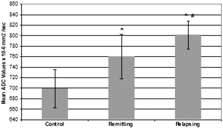
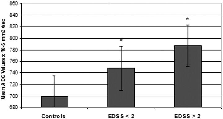
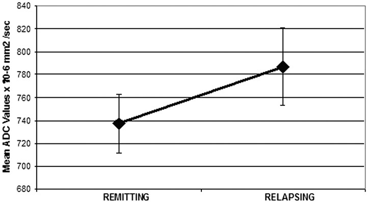
Results: Mean ADC values showed significant differences when relapsing, remitting, and control patients were compared. The relapsing or remitting phase showed significant difference when compared both with controls (P < .01) and between them (P < .05). Comparing mean ADC values of patients with clinical disability (EDSS <2 versus EDSS >/=2) also provided significant differences with the control group (P < .01). The data of patients showing a relapsing episode during the longitudinal part of the study showed a significant difference compared with data from their remitting phase (P < .01).
Conclusion: Brain microstructural changes can be detected and correlate with clinical impairment during the stages of MS. These changes modify with time in the relapsing group.
Figures
References
-
- Filippi M. Magnetization transfer imaging to monitor the evolution of individual multiple sclerosis lesions. Neurology 1999;53(Suppl 3):S18–22 - PubMed
-
- Filippi M, Campi A, Dousset V, et al. A magnetization transfer imaging study of normal-appearing white matter in multiple sclerosis. Neurology 1995;45:478–82 - PubMed
-
- Filippi M, Arnold DL, Comi G. Magnetic Resonance Spectroscopy in Multiple Sclerosis. Milan, Italy: Springer-Verlag;2001
MeSH terms
LinkOut - more resources
Full Text Sources