. 2007 May;13(5):763-80.
doi: 10.1261/rna.261807. Epub 2007 Mar 12.

A U-turn motif-containing stem-loop in the coronavirus 5' untranslated region plays a functional role in replication

Affiliations

A U-turn motif-containing stem-loop in the coronavirus 5' untranslated region plays a functional role in replication

Pinghua Liu et al. RNA. 2007 May.

Abstract

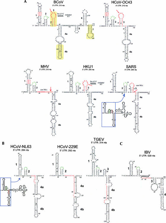
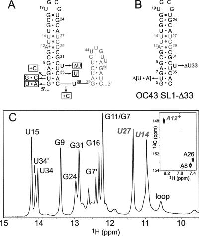
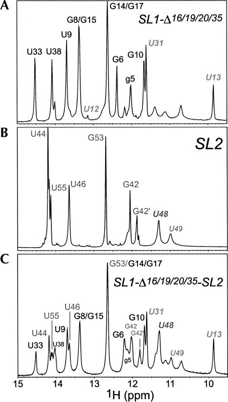
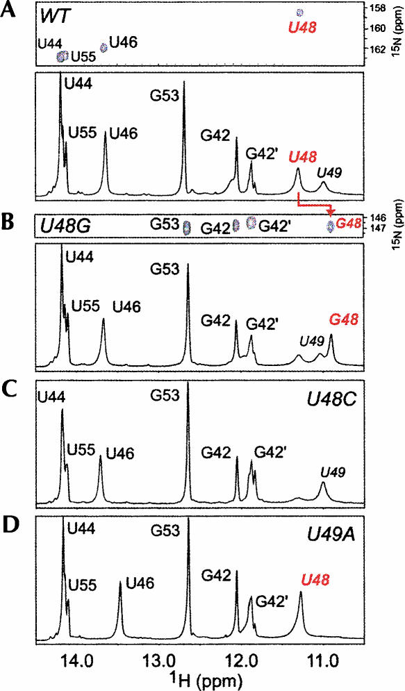
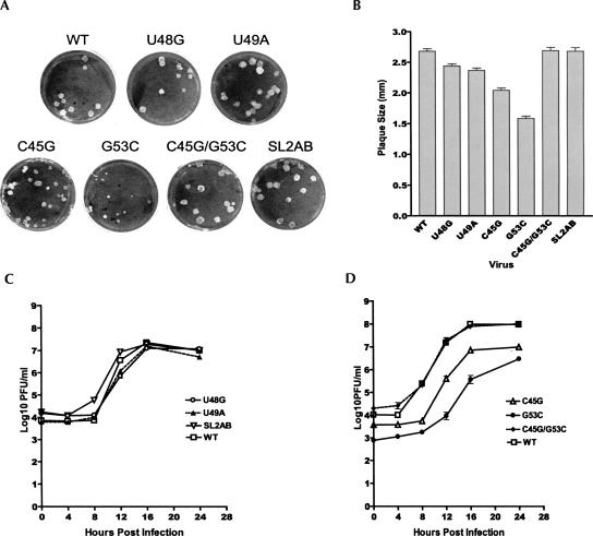
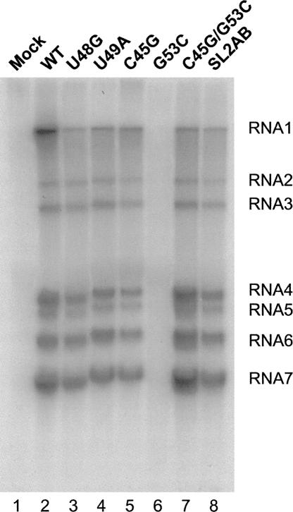
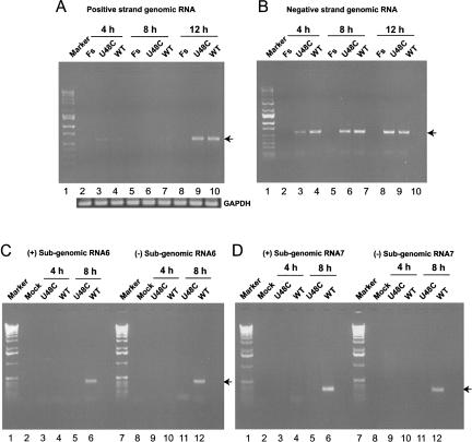
The 5' untranslated region (UTR) of the mouse hepatitis virus (MHV) genome contains cis-acting sequences necessary for transcription and replication. A consensus secondary structural model of the 5' 140 nucleotides of the 5' UTRs of nine coronaviruses (CoVs) derived from all three major CoV groups is presented and characterized by three major stem-loops, SL1, SL2, and SL4. NMR spectroscopy provides structural support for SL1 and SL2 in three group 2 CoVs, including MHV, BCoV, and HCoV-OC43. SL2 is conserved in all CoVs, typically containing a pentaloop (C47-U48-U49-G50-U51 in MHV) stacked on a 5 base-pair stem, with some sequences containing an additional U 3' to U51; SL2 therefore possesses sequence features consistent with a U-turn-like conformation. The imino protons of U48 in the wild-type RNA, and G48 in the U48G SL2 mutant RNA, are significantly protected from exchange with solvent, consistent with a hydrogen bonding interaction critical to the hairpin loop architecture. SL2 is required for MHV replication; MHV genomes containing point substitutions predicted to perturb the SL2 structure (U48C, U48A) were not viable, while those that maintain the structure (U48G and U49A) were viable. The U48C MHV mutant supports both positive- and negative-sense genome-sized RNA synthesis, but fails to direct the synthesis of positive- or negative-sense subgenomic RNAs. These data support the existence of the SL2 in our models, and further suggest a critical role in coronavirus replication.


Figures

FIGURE 1.
FIGURE 1.
(A) Predicted secondary structure models for the entire 5′ UTR of BCoV compared with the 5′ 140 nt of selected group 2 coronaviruses. (B) Predicted secondary structure models for three group 1 coronaviruses. (C) Predicted secondary structure model of a group 3 coronavirus, avian infectious bronchitis virus (IBV). (Bold numbers) Predicted stem–loops SL1, SL2, SL3, and SL4 (4a and 4b), (bold red letters) leader TRS-L sequences, (yellow) SL-II, SL-III, and SL-IV of Raman et al. (2003) and Raman and Brian (2005). Nucleotide positions are numbered according to GenBank accession numbers (BCoV-LUN: AF391542; HCoV-OC43: NC_005147; MHV-A59: NC_001846; HKU1: NC_006577; SARS-CoV: NC_004718; HCoV-NL63: NC_005831; HCoV-229E: NC_002645; TGEV: NC_002306); IBV: NC_001451). All models except one represent mfe structures, and are predicted by Mfold, PKNOTS, and ViennaRNA. The lone exception is MHV, which represents a structure within 1.7 kcal mol−1 of the mfe structure. If SL2, the strongest secondary structure in a covariation analysis (not shown), is forced to pair as indicated, the structure shown represents the mfe structure.
FIGURE 2.
FIGURE 2.
(A) Predicted secondary structures of SL1 and SL2 of HCoV-OC43. Nucleotide substitutions, insertions, and deletions at the base of SL1 that correspond to the BCoV-Lun sequence are indicated in the adjacent boxes. (B) HCoV-OC43 SL1 construct used for NMR studies, denoted OC43 SL1-Δ33. The U6-A36 base pair was excised to enable transcription by T7 RNA polymerase; the extrahelical U33 was also deleted. (C) Imino proton region of a 1D jump–return echo spectrum acquired at 10°C, pH 6.0 for OC43 SL1-Δ33. Resonance assignments were obtained from analysis of a homonuclear Watergate NOESY spectrum (τm=150 msec). The U14-U27 base pair was verified by the presence of a strong crosspeak in a NOESY spectrum acquired at a short mixing time (τm=50 msec). (Inset) Region of a natural-abundance 1H-13C HSQC spectrum acquired for OC43 SL1-Δ33, with assigned adenosine 13C2-1H2 cross-peaks indicated.
FIGURE 3.
FIGURE 3.
(A) Predicted secondary structure of MHV-A59 SL1. (B) Representation of the SL1, SL2 (boxed regions), and SL1-SL2 chimeras characterized in this study. All MHV SL1 constructs have a nonnative g5-c40 base pair (native sequence shown in brackets) at the base of SL1 to facilitate transcription by T7 RNA polymerase, and incorporate a nonnative base G-C pair base of SL2 and invert the MHV A44-U54 pair in MHV to the U-A pair present in SARS-coronavirus (the native MHV sequence is shown in brackets, see text for details). Imino proton regions of 1D jump–return echo spectra acquired at 10°C unless otherwise indicated for SL1-Δ16/19/20/35 (C), SL1-Δ16/19/20 (D), and SL1-Δ19/20 (E) (5°C). Resonance assignments were obtained from analysis of a homonuclear Watergate NOESY spectra (τm=150 msec) acquired for each RNA (300 msec for SL1-Δ19/20). Imino resonances for the U13-U31 base pair are indicated. (*) 14.2 ppm, the expected absence of the imino resonance for the A7-U38 base pair due to substitution of a nonnative g7-c38 base pair in this construct.
FIGURE 4.
FIGURE 4.
Imino proton regions of 1D jump–return echo spectra acquired at 10°C, pH 6.0 for SL1-Δ16/19/20/35 (A), SL2 (B), and SL1-Δ16/19/20/35-SL2 (C) RNA. See Fig. 3B for sequences for these RNA constructs. Note that the spectra for SL2 are characterized by slow conformational heterogeneity at the base of SL2 (G42-C56 and A43-U55 base pairs). The imino protons corresponding to U48 and U49 in the SL2 pentaloop are also indicated. The assignment of U12 is based on a weak NOE to a C32 amino proton.
FIGURE 5.
FIGURE 5.
Imino proton regions of 1D jump–return echo spectra acquired at 10°C, pH 6.0 for SL2 variants, with 1H-15N-HSQC spectra shown for 13C,15N-[U]-labeled WT SL2 and 13C,15N-[G]-labeled U48G SL2, as well. (A) WT SL2 (see Fig. 3B); (B) U48G SL2; (C) U48C SL2; and (D) U49A SL2. G42 and G42′ represent alternative conformations for the terminal G42-C56 base pair. The immediately adjacent U55 resonance is also doubled, indicative of heterogeneity at the base of SL2.
FIGURE 6.
FIGURE 6.
Growth phenotypes of SL2 mutant viruses. (A) Plaque morphologies of mutant and wild-type viruses. (B) Averaged plaque size of mutant and wild-type viruses. Plaque sizes were measured after DBT cell monolayers were stained with crystal violet. Plaque sizes were determined as described previously (Johnson et al. 2005). (C) One-step growth curves of viable loop and stem clustered point mutants. (D) One-step growth curve of stem point and compensatory mutations. Triplicate wells of DBT cells in 96-well plates were infected with mutant or wild-type viruses at a MOI of 3 and harvested at 0, 4, 8, 12, 16, and 24 h post-infection. Virus titers were determined by plaque assays. Error bars represent the standard errors of the mean.
FIGURE 7.
FIGURE 7.
Analysis of MHV specific RNAs synthesis. DBT cells infected with wild-type and mutant viruses RNAs were metabolically labeled from 6–12 h post-infection in the presence actinomycin D. Total RNAs were extracted and resolved on a formaldehyde agarose gel. (Lane 1) Mock infected cells, (lanes 28) cells infected with wild-type MHV A59–1000 (100%), U48G (72%), U49A (88%), C45G (57%), G53C (<1%), C45G/G53C (109%), and SL2AB (94%) genomes, respectively. The total amount of RNA synthesized relative to wild-type MHV A59–1000 is indicated in parentheses. (RNA 1–7) MHV-specific RNA bands. Molar ratios of indicated RNAs are compiled in Table 1.
FIGURE 8.
FIGURE 8.
Analysis of genomic and subgenomic RNAs of nonviable mutant U48C. RNAs were extracted 4, 8, and 12 h after cells were electroporated with in vitro assembled and transcribed Fs, wild-type MHV A59–1000, or U48C genomes and analyzed by RT-PCR or nested RT-PCR. (A) Positive-strand genomic RNA synthesis. (Marker, lane 1) 1-Kb DNA ladder, (Fs) RdRp frameshift mutant. (B) Negative-strand genomic RNA synthesis, with lanes marked as in panel A. (C) Positive-strand (left) and negative-strand (right) synthesis of RNA6; (Mock) Mock-infected cells. (D) Positive-strand (left) and negative-strand (right) synthesis of RNA7, with lanes marked as in panel C.
FIGURE 9.
FIGURE 9.
RT-PCR analysis of negative-strand genomic RNA of nonviable stem clustered point mutations SL2A and SL2B. (A) Full-length negative-strand genomic RNA synthesis of SL2A versus wild-type A59–1000. (B) Full-length negative-strand genomic RNA synthesis of SL2B versus wild-type A59–1000.

References

    1. Antao, V.P., Lai, S.Y., Tinoco I., Jr A thermodynamic study of unusually stable RNA and DNA hairpins. Nucleic Acids Res. 1991;19:5901–5905. - PMC - PubMed
    1. Baker, S.C., Sheh, C.-K., Soe, L.H., Chang, M.-F., Vannier, D.M., Lai, M.M.C. Identification of a domain required for autoproteolytic cleavage of murine coronavirus gene A polyprotein. J. Virol. 1989;63:3693–3699. - PMC - PubMed
    1. Brian, D.A., Spaan, W.J.M. Recombination and coronavirus defective interfering RNAs. Semin. Virol. 1997;8:101–111. - PMC - PubMed
    1. Brierley, I., Dos Ramos, F.J. Programmed ribosomal frameshifting in HIV-1 and the SARS-CoV. Virus Res. 2006;119:29–42. - PMC - PubMed
    1. Cabello-Villegas, J., Tworowska, I., Nikonowicz, E.P. Metal ion stabilization of the U-turn of the A37 N6-dimethylallyl-modified anticodon stem–loop of Escherichia coli tRNAPhe. Biochemistry. 2004;43:55–66. - PubMed

Publication types

LinkOut - more resources