. 2007 Mar 12;176(6):749-55.
doi: 10.1083/jcb.200612047.

Chromosome breakage after G2 checkpoint release

Affiliations

Chromosome breakage after G2 checkpoint release

Dorothee Deckbar et al. J Cell Biol. .

Abstract

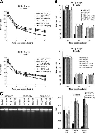
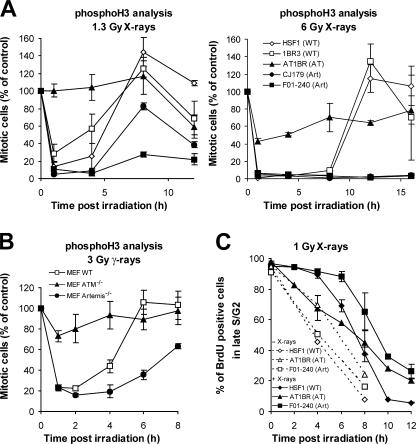
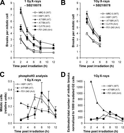
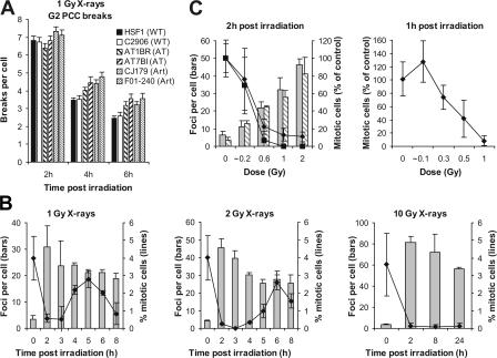
DNA double-strand break (DSB) repair and checkpoint control represent distinct mechanisms to reduce chromosomal instability. Ataxia telangiectasia (A-T) cells have checkpoint arrest and DSB repair defects. We examine the efficiency and interplay of ATM's G2 checkpoint and repair functions. Artemis cells manifest a repair defect identical and epistatic to A-T but show proficient checkpoint responses. Only a few G2 cells enter mitosis within 4 h after irradiation with 1 Gy but manifest multiple chromosome breaks. Most checkpoint-proficient cells arrest at the G2/M checkpoint, with the length of arrest being dependent on the repair capacity. Strikingly, cells released from checkpoint arrest display one to two chromosome breaks. This represents a major contribution to chromosome breakage. The presence of chromosome breaks in cells released from checkpoint arrest suggests that release occurs before the completion of DSB repair. Strikingly, we show that checkpoint release occurs at a point when approximately three to four premature chromosome condensation breaks and approximately 20 gammaH2AX foci remain.


Figures

Figure 1.
Figure 1.
A-T and Artemis primary human fibroblasts exhibit a DSB repair defect in G1 and G2. (A) γH2AX foci analysis in G1 and G2 phase cells after 1.5 Gy x-irradiation. Background foci numbers were ∼2 in G2 and 0.2 in G1. (B) γH2AX foci analysis in G1 and G2 phase cells after 1.5 Gy x-irradiation in the absence or presence of the ATM small molecule inhibitor KU55933 (ATMi). (C) FAR assay of [methyl-3H]thymidine-labeled exponentially growing cells irradiated in G2. (left) Ethidium bromide–stained PFGE gel from primary human fibroblasts irradiated with 10 Gy (for assessing DSB induction) or 80 Gy (for 48- and 72-h repair points) x-rays. The image has been grouped from different parts of the same gel for clarity. Note that the ethidium bromide signal represents DNA from all cells (G1, S, and G2) and is not used for evaluation. (right) FAR values calculated from the scintillation counts of gel slices derived from PFGE gels. Error bars indicate SEM.
Figure 2.
Figure 2.
Artemis cells show proficient checkpoint induction and a prolonged G2/M arrest. (A) PhosphoH3 analysis of primary human fibroblasts after 1.3 and 6 Gy x-irradiation. (B) PhosphoH3 analysis of MEFs after 3 Gy γ-irradiation. Data shown are the percentage of mitotic cells relative to unirradiated cells at time 0. (C) FACS analysis of BrdU-labeled primary human fibroblasts. The percentage of BrdU-positive cells in late S/G2 was assessed up to 12 h after 1 Gy x-irradiation given at 4 h after BrdU labeling (i.e., when BrdU-labeled cells have progressed into late S/G2). Dotted lines represent the percentage of BrdU-positive cells in late S/G2 without irradiation. Error bars indicate SEM.
Figure 3.
Figure 3.
ATM's repair and checkpoint functions contribute to prevent chromosome breakage. (A) Chromosome breaks per mitotic cell in metaphase spreads from primary human fibroblasts harvested at varying times after 1 Gy x-irradiation in the presence of aphidicolin. Breaks in unirradiated samples were <0.1 and were subtracted from the breaks in the irradiated samples. (B) Same analysis as in A in the presence of the Chk1/2 inhibitor SB218078. SB218078 did not cause chromosome breaks in unirradiated cells. (C) PhosphoH3 analysis of primary human fibroblasts after 1 Gy x-irradiation in the presence of aphidicolin (i.e., the same conditions used for the chromosomal analysis). The measured MIs were normalized to provide the same integral value of 1 for all three cell lines. (D) Estimation of the kinetics for total mitotic chromosome breakage. The values are derived from the number of chromosome breaks per mitotic cell for cells that enter mitosis at specific time points (taken from A) multiplied by the number of cells reaching mitosis at these times (taken from C). Error bars indicate SEM.
Figure 4.
Figure 4.
The G2/M checkpoint has a threshold of ∼3.5 PCC breaks and ∼20 γH2AX foci. (A) Analysis of G2 PCC chromosomal breaks in calyculin A–treated cells in the presence of aphidicolin at 2, 4, and 6 h after 1 Gy x-irradiation. Breaks in unirradiated samples were <0.2 and were subtracted from the breaks in the irradiated samples. P < 0.01 (t test) for AT1BR, AT7BI, CJ179, and F01-204 compared with HSF1 or C2906 at 4 and 6 h (but not at 2 h). (B) γH2AX analysis and mitotic counting of transformed MRC-5V1 fibroblasts at varying times after 1, 2, and 10 Gy x-irradiation in the presence of aphidicolin. The analysis was done on the same samples by counting the fraction of phosphoH3-positive mitotic cells and foci numbers in CENP-F–positive G2 cells, respectively. The pronounced decline in MI at 8 h after 1 and 2 Gy likely reflects the depletion of G2 cells. (C, left) Same analysis as in B evaluating transformed (MRC-5V1; gray bars, diamonds) and immortalized (48BR hTert; shaded bars, squares) fibroblasts 2 h after 0, 0.2, 0.6, 1, and 2 Gy x-irradiation. (right) PhosphoH3 analysis of primary (48BR) fibroblasts after 0, 0.1, 0.3, 0.5, and 1 Gy x-irradiation by FACS. γH2AX foci were scored on parallel samples and provided similar numbers to those of MRC-5V1 and 48BR hTert cells. Analysis was performed 1 h after IR, as pilot experiments showed that primary cells show a more rapid onset of checkpoint arrest. Note that very short exposure times were required to deliver the low doses in these experiments, resulting in potential errors in the estimated dosimetry. Error bars indicate SEM.

References

    1. Asakawa, Y., and E. Gotoh. 1997. A method for detecting sister chromatid exchanges using prematurely condensed chromosomes and immunogold-silver staining. Mutagenesis. 12:175–177. - PubMed
    1. Bekker-Jensen, S., C. Lukas, R. Kitagawa, F. Melander, M.B. Kastan, J. Bartek, and J. Lukas. 2006. Spatial organization of the mammalian genome surveillance machinery in response to DNA strand breaks. J. Cell Biol. 173:195–206. - PMC - PubMed
    1. Cedervall, B., R. Wong, N. Albright, J. Dynlacht, P. Lambin, and W.C. Dewey. 1995. Methods for the quantification of DNA double-strand breaks determined from the distribution of DNA fragment sizes measured by pulsed-field gel electrophoresis. Radiat. Res. 143:8–16. - PubMed
    1. Cornforth, M.N., and J.S. Bedford. 1985. On the nature of a defect in cells from individuals with ataxia-telangiectasia. Science. 227:1589–1591. - PubMed
    1. Cornforth, M.N., and J.S. Bedford. 1993. Ionizing radiation damage and its early development in chromosomes. In Advances in Radiation Biology, vol. 17. J.T. Lett and W.K. Sinclair, editors. Academic Press, San Diego. 423–496.

Publication types

MeSH terms