. 2007 Mar 12;176(6):779-86.
doi: 10.1083/jcb.200612080.

Requirement of ZO-1 for the formation of belt-like adherens junctions during epithelial cell polarization

Affiliations

Requirement of ZO-1 for the formation of belt-like adherens junctions during epithelial cell polarization

Junichi Ikenouchi et al. J Cell Biol. .

Abstract

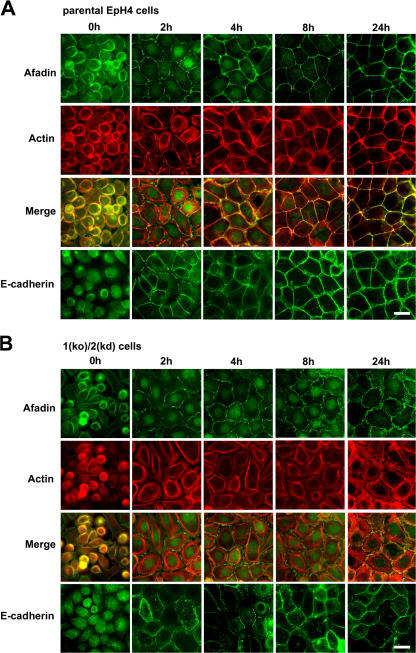
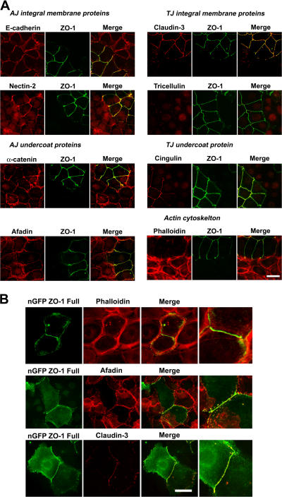
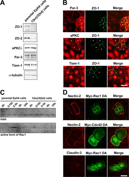
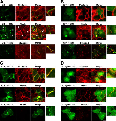
The molecular mechanisms of how primordial adherens junctions (AJs) evolve into spatially separated belt-like AJs and tight junctions (TJs) during epithelial polarization are not well understood. Previously, we reported the establishment of ZO-1/ZO-2-deficient cultured epithelial cells (1[ko]/2[kd] cells), which lacked TJs completely. In the present study, we found that the formation of belt-like AJs was significantly delayed in 1(ko)/2(kd) cells during epithelial polarization. The activation of Rac1 upon primordial AJ formation is severely impaired in 1(ko)/2(kd) cells. Our data indicate that ZO-1 plays crucial roles not only in TJ formation, but also in the conversion from "fibroblastic" AJs to belt-like "polarized epithelial" AJs through Rac1 activation. Furthermore, to examine whether ZO-1 itself mediate belt-like AJ and TJ formation, respectively, we performed a mutational analysis of ZO-1. The requirement for ZO-1 differs between belt-like AJ and TJ formation. We propose that ZO-1 is directly involved in the establishment of two distinct junctional domains, belt-like AJs and TJs, during epithelial polarization.


Figures

Figure 1.
Figure 1.
Retardation of the formation of belt-like AJs and linear actin cables in 1(ko)/2(kd) cells during epithelial polarization. Parental EpH4 cells (A) and 1(ko)/2(kd) cells (B) were cultured overnight in low Ca2+ medium overnight, and their polarization was initiated by transferring to normal Ca2+ medium. After a 0, 2-, 4-, 8-, or 24-h incubation, cells were fixed and stained with anti-afadin mAb, anti-E cadherin mAb, and phalloidin. Bars, 10 μm.
Figure 2.
Figure 2.
Molecular assembly of junctions in 1(ko)/2(kd) cells. (A) Parental EpH4 cells and 1(ko)/2(kd) cells were cocultured and doubly stained with the indicated antibodies 24 h after relpating. Bar, 10 μm. (B) The retardation of the formation of belt-like AJs, TJs, and linear actin cables were canceled by the exogenous expression of GFP-tagged full-length ZO-1. Cells were fixed and double stained with the indicated antibodies 24 h after transfection. Bar, 15 μm.
Figure 3.
Figure 3.
Impaired activation of Rac1 in 1(ko)/2(kd) cells during epithelial polarization. (A) Immunoblotting of whole-cell lysates of parental EpH4cells and 1(ko)/2(kd) cells with the indicated antibodies. (B) Parental EpH4 cells and 1(ko)/2(kd) cells were cocultured and doubly stained with the indicated antibodies 24 h after replating. Parental cells are indicated by asterisks. (C) Rac activity assays from parental EpH4 cells and 1(ko)/2(kd) cells after a Ca2+ switch for the indicated times. (D) 1(ko)/2(kd) cells were transiently transfected with myc-Rac1-DA or Cdc42-DA. Cells were double stained for myc and anti–nectin -2 antibody (as an AJ marker) or anti–claudin-3 antibody (as a TJ marker) 24 h after transfection. Bars, 10 μm.
Figure 4.
Figure 4.
Schematic representation of ZO-1 deletion mutants. (A) Schematic drawings of deletion constructs of ZO-1. Amino acid residues of ZO-1 are shown in parentheses. (B) Total cell lysates derived from HEK293 cells transiently expressing each construct were separated by SDS-PAGE and immunoblotted with anti-GFP pAb. (C) Stable EpH4 lines expressing the deletion constructs of ZO-1 were stained with antibodies against GFP and claudin-3. Note that ZO-11-871, ZO-1516-1746, and ZO-1804-1746 were incorporated into TJs and did not affect the localization of claudin-3, whereas ZO-11-805 was distributed along the lateral plasma membrane and induced ectopic TJs. Bar, 10 μm.
Figure 5.
Figure 5.
Rescuing belt-like AJ and/or TJ formation by deletion constructs of ZO-1 in 1(ko)/2(kd) cells. 1(ko)/2(kd) cells were transfected with GFP-tagged ZO-11-805 (A), ZO-11-871 (B), ZO-1516-1746 (C), and ZO-1804-1746 (D). Cells were fixed and stained with the indicated antibodies 24 h after transfection. The formation of belt-like AJs was not observed at cell–cell contacts between ZO-11-805 expressing 1(ko)/2(kd) cells (arrowheads). Bar, 10 μm.

References

    1. Anderson, J.M., C.M. Van Itallie, and A.S. Fanning. 2004. Setting up a selective barrier at the apical junction complex. Curr. Opin. Cell Biol. 16:140–145. - PubMed
    1. Ando-Akatsuka, Y., S. Yonemura, M. Itoh, M. Furuse, and S. Tsukita. 1999. Differential behavior of E-cadherin and occludin in their colocalization with ZO-1 during the establishment of epithelial cell polarity. J. Cell. Physiol. 179:115–125. - PubMed
    1. Chen, X., and I.G. Macara. 2005. Par-3 controls tight junction assembly through the Rac exchange factor Tiam1. Nat. Cell Biol. 7:262–269. - PubMed
    1. Furuse, M., and S. Tsukita. 2006. Claudins in occluding junctions of humans and flies. Trends Cell Biol. 16:181–188. - PubMed
    1. Gonzalez-Mariscal, L., A. Betanzos, and A. Avila-Flores. 2000. MAGUK proteins: structure and role in the tight junction. Semin. Cell Dev. Biol. 11:315–324. - PubMed

Publication types

MeSH terms