. 2007 Mar 12;176(6):787-93.
doi: 10.1083/jcb.200611044.

Absence of integrin-mediated TGFbeta1 activation in vivo recapitulates the phenotype of TGFbeta1-null mice

Affiliations

Absence of integrin-mediated TGFbeta1 activation in vivo recapitulates the phenotype of TGFbeta1-null mice

Zhiwei Yang et al. J Cell Biol. .

Abstract

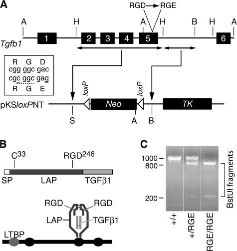
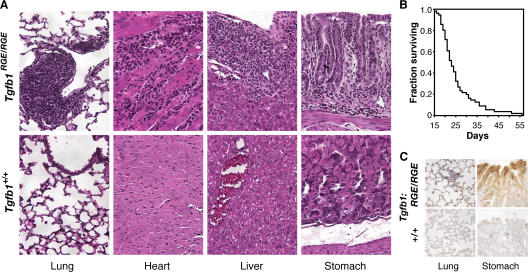
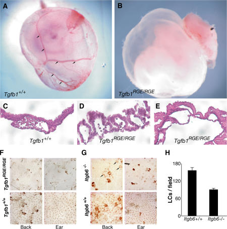
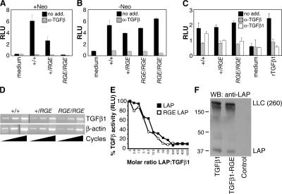
The multifunctional cytokine transforming growth factor (TGF) beta1 is secreted in a latent complex with its processed propeptide (latency-associated peptide [LAP]). TGFbeta1 must be functionally released from this complex before it can engage TGFbeta receptors. One mechanism of latent TGFbeta1 activation involves interaction of the integrins alpha v beta6 and alpha v beta8 with an RGD sequence in LAP; other putative latent TGFbeta1 activators include thrombospondin-1, oxidants, and various proteases. To assess the contribution of RGD-binding integrins to TGFbeta1 activation in vivo, we created a mutation in Tgfb1 encoding a nonfunctional variant of the RGD sequence (RGE). Mice with this mutation (Tgfb1(RGE/RGE)) display the major features of Tgfb1(-/-) mice (vasculogenesis defects, multiorgan inflammation, and lack of Langerhans cells) despite production of normal levels of latent TGFbeta1. These findings indicate that RGD-binding integrins are requisite latent TGFbeta1 activators during development and in the immune system.


Figures

Figure 1.
Figure 1.
Generation of mice with a targeted mutation of Tgfb1. (A) Schematic of Tgfb1 (top) and fragments thereof used for insertion in targeting vector (bottom). Box shows mutations (solid underlines) introduced in exon 5 to encode a BstUI restriction site (dashed underline) and the D-to-E mutation. Restriction sites: A, ApaI; B, BamHI; H, HindIII; S, SalI. Neo, neomycin resistance cassette; TK, thymidine kinase cassette. (B) The TGFβ1 mRNA encodes a protein sequence consisting of a signal peptide (SP), propeptide (LAP), and the TGFβ1 cytokine (top). The integrin-binding motif RGD is near the C terminus of LAP. The processed latent factor consists of noncovalently associated disulfide-linked homodimers of the LAP and TGFβ1 monomers (bottom). LAP can be disulfide linked via cys-33 to one of the cysteine-repeat domains (ovals) of LTBP-1, -3, or -4. (C) PCR genotyping results from Tgfb1+/+, Tgfb1+/RGE, and Tgfb1RGE/RGE mice.
Figure 2.
Figure 2.
Tgfb1RGE/RGE mice develop fatal multiorgan inflammation. (A) Histology of inflammatory lesions in lung, heart, liver, and stomach of Tgfb1RGE/RGE mice (hematoxylin and eosin staining). (B) Kaplan-Meier survival curve for Tgfb1RGE/RGE mice (n = 54). (C) Increased expression of β6 protein in stomach and lung epithelium of Tgfb1RGE/RGE mice.
Figure 3.
Figure 3.
Defects in vascular development and LCs in Tgfb1RGE/RGE mice. (A and B) Vasculature is present in wild-type yolk sac (arrows) but is not identifiable in an E12.5 Tgfb1RGE/RGE embryo. (C–E) Histologic appearance of control and Tgfb1RGE/RGE E12.5 yolk sacs. In the mutant yolk sacs, there is poor contact between endothelial and mesothelial layers and, in E, absence of blood cells. (F) LCs are absent in epidermis from back and ear of Tgfb1RGE/RGE mice. (G) LCs are less abundant in back epidermis (arrows) and absent in ear epidermis of Itgb6−/− mice. (H) Compared with control mice, Itgb6−/− mice have fewer LCs in back epidermis. P = 0.001. Error bars indicate means ± SEM.
Figure 4.
Figure 4.
The RGE mutation does not affect Tgfb1 gene expression, LAP inhibitory function, or latent TGFβ1 processing and secretion. Serum TGFβ1 levels in Tgfb1−/−, Tgfb1+/RGE, and Tgfb1RGE/RGE mice before (A) and after (B) removal of a Neo sequence within the mutated Tgfb1 allele. (C) TGFβ levels in medium conditioned by lung fibroblasts derived from Tgfb1−/−, Tgfb1+/RGE, and Tgfb1RGE/RGE mice. Effects of an anti-TGFβ antibody active against all three TGFβ isoforms (α-TGFβ) and of an antibody that inhibits only TGFβ1 (α-TGFβ1) are shown. Error bars indicate means ± SEM. (D) Tgfb1 gene expression was assessed by semiquantitative RT-PCR using RNA isolated from Tgfb1−/−, Tgfb1+/RGE, and Tgfb1RGE/RGE lung fibroblasts. Reverse-transcribed β-actin mRNA was amplified as a control. (E) Recombinant wild-type and RGE mutant LAP inhibit equally the activity of recombinant TGFβ1, measured with a cell-based luciferase assay. Luciferase activity, expressed as relative light units (RLU), is proportional to TGFβ activity. (F) Wild-type and RGE mutant TGFβ1 cDNAs were expressed in LTBP1S-expressing CHO cells. Secreted proteins were separated by SDS-PAGE in nonreducing conditions and probed with an anti-LAP antibody. Large latent complex (LLC) indicates migration of the LAP–LTBP1 complex.

References

    1. Abe, M., J.G. Harpel, C.N. Metz, I. Nunes, D.J. Loskutoff, and D.B. Rifkin. 1994. An assay for transforming growth factor-β using cells transfected with a plasminogen activator inhibitor-1 promoter-luciferase construct. Anal. Biochem. 216:276–284. - PubMed
    1. Annes, J.P., D.B. Rifkin, and J.S. Munger. 2002. The integrin αvβ6 binds and activates latent TGFβ3. FEBS Lett. 511:65–68. - PubMed
    1. Annes, J.P., J.S. Munger, and D.B. Rifkin. 2003. Making sense of latent TGFβ activation. J. Cell Sci. 116:217–224. - PubMed
    1. Annes, J.P., Y. Chen, J.S. Munger, and D.B. Rifkin. 2004. Integrin αvβ6-mediated activation of latent TGF-β requires the latent TGF-β binding protein-1. J. Cell Biol. 165:723–734. - PMC - PubMed
    1. Araya, J., S. Cambier, A. Morris, W. Finkbeiner, and S.L. Nishimura. 2006. Integrin-mediated transforming growth factor-β activation regulates homeostasis of the pulmonary epithelial-mesenchymal trophic unit. Am. J. Pathol. 169:405–415. - PMC - PubMed

Publication types