. 2007 Mar 19;204(3):481-7.
doi: 10.1084/jem.20061791. Epub 2007 Mar 12.

IL-31-IL-31R interactions negatively regulate type 2 inflammation in the lung

Affiliations

IL-31-IL-31R interactions negatively regulate type 2 inflammation in the lung

Jacqueline G Perrigoue et al. J Exp Med. .

Abstract

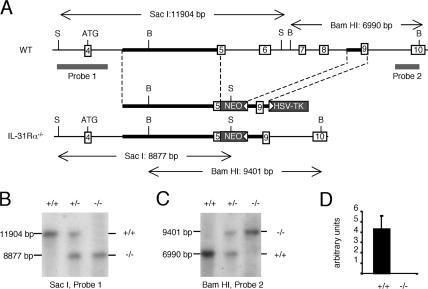
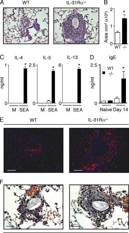
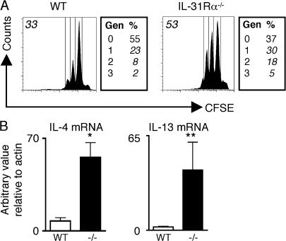
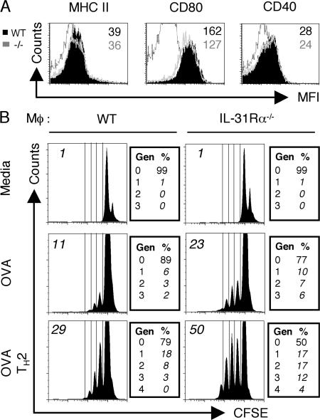
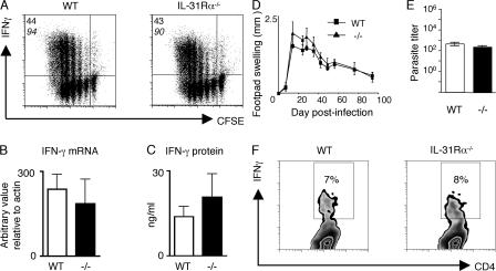
Interleukin (IL) 31Ralpha (glycoprotein 130-like monocyte receptor and glycoprotein 130-like receptor) heterodimerizes with oncostatin M receptor beta to bind IL-31, a cytokine expressed preferentially by CD4(+) T helper type 2 (Th2) cells. However, the functions of IL-31-IL-31R signaling in immune regulation remain unknown. Here, we identify a novel role for IL-31R in limiting type 2 inflammation in the lung. After intravenous injection of Schistosoma mansoni eggs, IL-31Ralpha(-/-) mice developed severe pulmonary inflammation, characterized by an increase in the area of granulomatous inflammation, increased numbers of resistin-like molecule alpha(+) cells, and enhanced collagen deposition compared to WT counterparts. In vitro, macrophages generated from IL-31Ralpha(-/-) mice promoted enhanced ovalbumin-specific CD4(+) T cell proliferation and purified naive CD4(+) T cells from IL-31Ralpha(-/-) mice exhibited enhanced proliferation and expression of Th2 cytokines, identifying a T cell- and macrophage-intrinsic regulatory function for IL-31R signaling. In contrast, the generation of CD4(+) T cell-mediated Th1 responses were normal in IL-31Ralpha(-/-) mice, suggesting that the regulatory role of IL-31R signaling is limited to type 2 responses. Together, these data implicate IL-31R signaling as a novel negative regulatory pathway that specifically limits type 2 inflammation.


Figures

Figure 1.
Figure 1.
Generation of IL-31Rα−/− mice. (A) The relevant section of the native IL-31Rα locus (top), the targeting construct (middle), and the correctly targeted locus (bottom) are depicted. Open boxes with numbers indicate exons and gray boxes indicate the selection genes neomycin phosphotransferase (NEO) and thymidine kinase (TK), with white arrowheads depicting their transcriptional orientation. Thick black lines indicate the position of the 5′ and 3′ homology arms. The size of restriction fragments resulting from digestion with SacI (S) and BamHI (B) are indicated, and the location of the two probes used to detect these fragments by Southern blot are shown by thick gray lines. (B and C) Southern blot analysis of SacI digests probed with probe 1 and BamHI digests probed with probe II, respectively. Genotype is indicated as follows: +/+, WT; +/−, heterozygous; −/−, IL-31Rα−/−. (D) Expression of IL-31Rα relative to rpl19 was determined by quantitative Taqman PCR.
Figure 2.
Figure 2.
Enhanced S. mansoni egg–induced type 2 inflammation in the lungs of IL-31Rα−/− mice. (A and B) Paraffin sections of lungs from egg-injected mice at day 14 after injection were H &E stained and the area of inflammation surrounding WT (open bar) and IL-31Rα−/− (closed bar) S. mansoni egg-induced granulomas was measured using OpenLab software. Mean ± SEM; n = 40 granulomas per group. (C) Draining mediastinal lymph node cells from egg-injected WT and IL-31Rα−/− mice were stimulated with either media or 20 μg/ml S. mansoni egg antigen (SEA) for 48 h. Supernatants were analyzed by ELISA for IL-4, -5, and -13. Mean ± SEM of replicate cultures. (D) Serum levels of IgE from WT and IL-31Rα−/− mice were measured by ELISA. Mean ± SEM; n = 3 mice per group. (E) Immunofluorescence staining for RELM-α (red) and counterstain for DAPI (blue). (F) Collagen deposition in the lungs of WT and IL-31Rα−/− mice was detected by Masson's trichrome staining; blue staining demarks collagen. Results are representative of three independent experiments of three mice per group. Asterisk indicates statistically significant as determined by two-tailed Student's t test (P < 0.05). Bars, 60 μm.
Figure 3.
Figure 3.
Naive IL-31Rα−/− CD4+ T cells exhibit enhanced proliferation and expression of Th2 effector cytokines. (A) Purified naive CD4+ T cells from WT or IL-31Rα−/− mice were CFSE labeled and stimulated with plate-bound anti-CD3/anti-CD28 for 4 d under neutral conditions. Boxes to the right indicate the percentage of events in each proliferating generation (Gen). Numbers in italics in upper left indicate percentage of CFSE-dim cells. (B) cDNA derived from stimulated cells was assayed by real-time PCR for IL-4 or IL-13 mRNA levels relative to actin. Results expressed as the mean ± SEM for three independent experiments. *, P < 0.05; **, P < 0.1.
Figure 4.
Figure 4.
IL-31Rα−/− macrophages exhibit enhanced accessory cell function. (A) Bone marrow–derived macrophages from either WT (filled black) or IL-31Rα−/− (gray line) mice were characterized for surface molecule expression of MHC II, CD80, or CD40. Numbers refer to mean fluorescence intensity (black, WT; gray, IL-31Rα−/−). Black line is isotype control. (B) WT or IL-31Rα−/− macrophages were pulsed overnight with OVA protein in the presence or absence of IL-4 when indicated. CFSE-labeled OTII T cells were added 24 h after OVA pulse and co-cultured with macrophages for 4 d under neutral or Th2-polarizing conditions. Histograms are gated on the live CD4+ population; boxes to the right indicate the percentage of events in each proliferating generation (Gen). Numbers in italics in top left indicate percentage of CFSE-dim cells. Data are representative of six independent experiments.
Figure 5.
Figure 5.
Normal Th1 cell differentiation in IL-31Rα−/− mice. (A) Purified CD4+ T cells from WT and IL-31Rα−/− mice were CFSE labeled and stimulated with plate-bound anti-CD3/anti-CD28 under Th1-polarizing conditions. Cells were assayed for proliferation and cytokine production by flow cytometry. The top number indicates the percentage of proliferating CD4+ T cells producing IFN-γ and the bottom number in italics is the percentage of CFSE-dim cells. (B) cDNA from purified naive CD4+ T cells cultured under Th1-polarizing conditions was subjected to real-time PCR analysis for IFN-γ expression relative to actin. (C) Supernatants from the cultures in A were assayed by ELISA for IFN-γ levels. (D) WT and IL-31Rα−/− mice were infected with L. major promastigotes in the hind footpad. Lesion size was determined by measuring swelling of the infected footpad and subtracting that of the uninfected contralateral footpad. (E) Parasite load in the footpad was quantified by limiting dilution analysis at 12 wk after infection. Values represent the mean ± SEM for three mice per group. (F) Cells from the draining lymph nodes of L. major– infected WT or IL-31Rα−/− mice were restimulated with soluble L. major antigen and analyzed by flow cytometry. Plots are gated on CD4+ cells and the number in the box indicates the percentage of CD4+ T cells producing IFN-γ.

References

    1. Boulay, J.L., J.J. O'Shea, and W.E. Paul. 2003. Molecular phylogeny within type I cytokines and their cognate receptors. Immunity. 19:159– 163. - PubMed
    1. Hofmann, S.R., R. Ettinger, Y.J. Zhou, M. Gadina, P. Lipsky, R. Siegel, F. Candotti, and J.J. O'Shea. 2002. Cytokines and their role in lymphoid development, differentiation and homeostasis. Curr. Opin. Allergy Clin. Immunol. 2:495–506. - PubMed
    1. Hunter, C.A. 2005. New IL-12-family members: IL-23 and IL-27, cytokines with divergent functions. Nat. Rev. Immunol. 5:521–531. - PubMed
    1. Dreuw, A., S. Radtke, S. Pflanz, B.E. Lippok, P.C. Heinrich, and H.M. Hermanns. 2004. Characterization of the signaling capacities of the novel gp130-like cytokine receptor. J. Biol. Chem. 279:36112–36120. - PubMed
    1. Diveu, C., E. Lelievre, D. Perret, A.H. Lak-Hal, J. Froger, C. Guillet, S. Chevalier, F. Rousseau, A. Wesa, L. Preisser, et al. 2003. GPL, a novel cytokine receptor related to GP130 and leukemia inhibitory factor receptor. J. Biol. Chem. 278:49850–49859. - PubMed

Publication types

MeSH terms