. 2007 Mar 19;204(3):633-43.
doi: 10.1084/jem.20062041. Epub 2007 Mar 12.

Aberrant immunoglobulin class switch recombination and switch translocations in activated B cell-like diffuse large B cell lymphoma

Affiliations

Aberrant immunoglobulin class switch recombination and switch translocations in activated B cell-like diffuse large B cell lymphoma

Georg Lenz et al. J Exp Med. .

Abstract

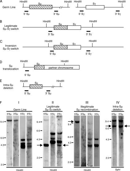
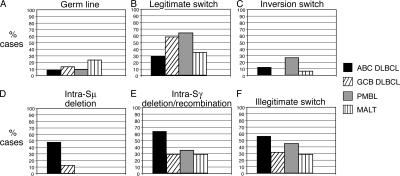
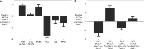
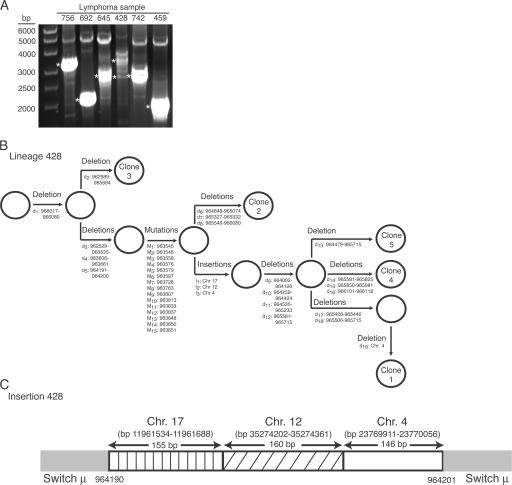
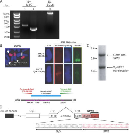
To elucidate the mechanisms underlying chromosomal translocations in diffuse large B cell lymphoma (DLBCL), we investigated the nature and extent of immunoglobulin class switch recombination (CSR) in these tumors. We used Southern blotting to detect legitimate and illegitimate CSR events in tumor samples of the activated B cell-like (ABC), germinal center B cell-like (GCB), and primary mediastinal B cell lymphoma (PMBL) subgroups of DLBCL. The frequency of legitimate CSR was lower in ABC DLBCL than in GCB DLBCL and PMBL. In contrast, ABC DLBCL had a higher frequency of internal deletions within the switch mu (Smu) region compared with GCB DLBCL and PMBL. ABC DLBCLs also had frequent deletions within Sgamma and other illegitimate switch recombinations. Sequence analysis revealed ongoing Smu deletions within ABC DLBCL tumor clones, which were accompanied by ongoing duplications and activation-induced cytidine deaminase-dependent somatic mutations. Unexpectedly, short fragments derived from multiple chromosomes were interspersed within Smu in one case. These findings suggest that ABC DLBCLs have abnormalities in the regulation of CSR that could predispose to chromosomal translocations. Accordingly, aberrant switch recombination was responsible for translocations in ABC DLBCLs involving BCL6, MYC, and a novel translocation partner, SPIB.


Figures

Figure 1.
Figure 1.
Analysis of class switch recombination by Southern blot. (A–E) The position of HindIII restriction sites are shown. Thick lines indicate binding sites of respective probes. (A) Germ line configuration of switch μ and switch γ regions. (B) Legitimate switch μ to γ, detected by 5′ Sμ and 3′ Sγ probe. (C) Inversion switch μ to γ, detected by 5′ Sμ and 5′ Sγ probe. (D) Chromosomal translocation, detected by 5′ Sμ probe. (E) Intra-switch μ deletion, detected by 5′ Sμ and 3′ Sμ probe. (F) Applied restriction enzyme is indicated below lanes. The probes used for hybridization are shown above each lane. Panel I: Restriction digest of placental DNA shows germ line configuration. Panel II: Restriction digest of patient sample 529 genomic DNA. Arrows indicate legitimate switch recombination μ to γ. Panel III: Restriction digest of SUDHL-2 genomic DNA. Arrow indicates illegitimate switch recombination detected by 5′ Sμ probe. Panel IV: Restriction digest of patient sample 1002 genomic DNA. Arrows indicate intra-switch μ deletion.
Figure 2.
Figure 2.
Summary of the analysis of class switch recombination in 92 DLBCL and 17 MALT lymphoma patients. (A) Germ line. No significant differences can be observed between the different lymphoma subtypes. (B) Legitimate switch recombination. GCB DLBCL and PMBL show a significantly higher frequency of legitimate switch recombinations compared with ABC DLBCL (ABC DLBCL vs. GCB DLBCL, P = 0.012; ABC DLBCL vs. PMBL, P = 0.035). (C) Inversion switch recombination. ABC DLBCL shows a significantly higher rate of inversion switch recombinations compared with GCB DLBCL (ABC DLBCL vs. GCB DLBCL, P = 0.045). (D) Intra-switch μ deletions. ABC DLBCL shows a significantly higher rate of intra-switch μ deletions compared with GCB DLBCL, PMBL, or MALT (ABC DLBCL vs. GCB DLBCL, P = 0.0012; ABC DLBCL vs. PMBL, P = 0.003; ABC DLBCL vs. MALT, P = 0.0004). (E) Intra-switch γ deletion/recombination. ABC DLBCL shows a significantly higher rate of intra-switch γ deletion/recombination compared with GCB DLBCL and MALT (ABC DLBCL vs. GCB DLBCL, P = 0.002; ABC DLBCL vs. MALT, P = 0.013). (F) Illegitimate switch recombination. ABC DLBCL shows a significantly higher rate of illegitimate switch recombinations compared with GCB DLBCL (ABC DLBCL vs. GCB DLBCL, P = 0.037).
Figure 3.
Figure 3.
Relative AID mRNA expression in different lymphoma subtypes. (A) ABC DLBCLs and PMBLs express the highest level of AID mRNA. (B) Comparison of AID mRNA expression in ABC DLBCL and GCB DLBCL germ line cases versus non-germ line cases. Non-germ line cases express higher AID mRNA compared with germ line cases.
Figure 4.
Figure 4.
Analysis of intra-switch μ deletions. (A) PCR amplification of intra-switch μ deletions (ethidium bromide–stained 0.8% agarose gel). Asterisks indicate cloned PCR bands for sequence analysis. (B) Lineage model of aberrations in ABC DLBCL patient sample 428. d, deletion; M, mutation; I, insertion. Open circles represent putative clonal precursors. Numbers refer to GenBank accession number NG_001019.4. (C) Sequence analysis of DNA insertions into Sμ in ABC DLBCL case 428. bp numbers refer to accession number Hs17_10875 for chromosome 17, accession number Hs12_29578 for chromosome 12, and accession number Hs4_16510 for chromosome 4.
Figure 5.
Figure 5.
Switch translocations in DLBCL. (A) PCR for detection of BCL6/MYC switch translocations (ethidium bromide–stained 0.8% agarose gel). Lane 1, Sγ-MYC translocation in ABC DLBCL case 1000; lane 2, Sγ-MYC translocation in GCB DLBCL cell line NUDUL-1; lane 3, Sμ-BCL6 translocation in ABC DLBCL case 709. (B) Whole chromosome painting of chromosome 19 detects t(14; 19) and t(19; 22; 4; 18). Hybridization of t(14; 19) with spanning and telomeric BAC SPIB probes. No hybridization with centromeric BAC probe. Hybridization of t(19; 22; 4; 18) with centromeric and spanning BAC SPIB probe, no hybridization with telomeric probe. (C) Detection of switch γ-SPIB translocation by Southern blot in OCI-Ly3. (D) Sequence analysis of switch γ-SPIB translocation. Δ, deletion in Sγ.

References

    1. 1997. A clinical evaluation of the International Lymphoma Study Group classification of non-Hodgkin's lymphoma. The Non-Hodgkin's Lymphoma Classification Project. Blood. 89:3909–3918. - PubMed
    1. Coiffier, B. 200 1. Diffuse large cell lymphoma. Curr. Opin. Oncol. 13:325–334. - PubMed
    1. Alizadeh, A.A., M.B. Eisen, R.E. Davis, C. Ma, I.S. Lossos, A. Rosenwald, J.C. Boldrick, H. Sabet, T. Tran, X. Yu, et al. 2000. Distinct types of diffuse large B-cell lymphoma identified by gene expression profiling. Nature. 403:503–511. - PubMed
    1. Rosenwald, A., G. Wright, W.C. Chan, J.M. Connors, E. Campo, R.I. Fisher, R.D. Gascoyne, H.K. Muller-Hermelink, E.B. Smeland, J.M. Giltnane, et al. 2002. The use of molecular profiling to predict survival after chemotherapy for diffuse large-B-cell lymphoma. N. Engl. J. Med. 346:1937–1947. - PubMed
    1. Rosenwald, A., G. Wright, K. Leroy, X. Yu, P. Gaulard, R.D. Gascoyne, W.C. Chan, T. Zhao, C. Haioun, T.C. Greiner, et al. 2003. Molecular diagnosis of primary mediastinal B cell lymphoma identifies a clinically favorable subgroup of diffuse large B cell lymphoma related to Hodgkin lymphoma. J. Exp. Med. 198:851–862. - PMC - PubMed

Publication types

MeSH terms