. 2007 Mar 19;204(3):475-80.
doi: 10.1084/jem.20062465. Epub 2007 Mar 12.

Lack of Foxp3 function and expression in the thymic epithelium

Affiliations

Lack of Foxp3 function and expression in the thymic epithelium

Adrian Liston et al. J Exp Med. .

Abstract

Foxp3 is essential for the commitment of differentiating thymocytes to the regulatory CD4(+) T (T reg) cell lineage. In humans and mice with a genetic Foxp3 deficiency, absence of this critical T reg cell population was suggested to be responsible for the severe autoimmune lesions. Recently, it has been proposed that in addition to T reg cells, Foxp3 is also expressed in thymic epithelial cells where it is involved in regulation of early thymocyte differentiation and is required to prevent autoimmunity. Here, we used genetic tools to demonstrate that the thymic epithelium does not express Foxp3. Furthermore, we formally showed that genetic abatement of Foxp3 in the hematopoietic compartment, i.e. in T cells, is both necessary and sufficient to induce the autoimmune lesions associated with Foxp3 loss. In contrast, deletion of a conditional Foxp3 allele in thymic epithelial cells did not result in detectable changes in thymocyte differentiation or pathology. Therefore, in mice the only known role for Foxp3 remains promotion of T reg cell differentiation within the T cell lineage, whereas there is no role for Foxp3 in thymic epithelial cells.


Figures

Figure 1.
Figure 1.
No expression of Foxp3 in the thymic epithelium. (A) Expression of GFP in CD45 thymic stroma from wild-type (shaded) and Foxp3GFP mice (line). Data is representative of four independent experiments. (B) Immunohistochemical analysis of Foxp3 expression in the thymus of Foxp3GFP, Foxp3GFPRag2 /, and Foxp3 Rag / mice. Thymic sections were stained with the affinity-purified rabbit anti-Foxp3 antibody. Bar, 100 μm.
Figure 2.
Figure 2.
Disruption of Foxp3 in the T cell lineage is necessary and sufficient to cause autoimmune syndrome. (A) No expression of CD4 in the CD45 thymic stroma. Left: expression of CD4 in wild-type CD45 thymic stroma. Thymic stromal cells were analyzed after incubation with anti-CD4 antibodies (solid line) or no antibody control (shaded). Right: expression of YFP in CD45 thymic stroma and CD45+ thymocytes from wild-type (shaded) and Rosa-stopflYFP × CD4-Cre (solid line) mice. Data representative of 11 and 3 experiments, respectively. (B) Expression of YFP from wild-type (shaded) and Rosa-stopflYFP × Foxn1Cre (line) mice in CD45G8.8+ epithelium, CD45G8.8 stroma, and thymocytes. Data is representative of three experiments. (C) Analysis of gross clinical signs and lung and skin histopathology in Foxp3fl × Foxn1Cre (n = 7; no histological infiltrates at 4 or 9 wk of age, no visible disease at >16 wk of age), Foxp3fl × CD4-Cre (n = 6; average life span 4 wk), Foxp3 (n = 7; average life span 4 wk), and wild-type or Foxp3fl mice (n = 9; no histological infiltrates or visible disease at >16 wk of age). Bar, 1 cm. For representative histology sections shown, lung and ear skin were taken from 4-wk-old male Foxp3fl, Foxp3fl CD4-Cre, and Foxp3fl Foxn1Cre mice. Arrows indicate inflammatory infiltrate. Bar, 100 μm. (D) Disease onset in neonatal Rag1 / mice reconstituted with Foxp3 scurfy nude BM (▪; n = 4) or Foxp3 wild-type nude BM (▴; n = 4), and neonatal Rag1 / Foxp3 scurfy mice reconstituted with Foxp3 scurfy nude BM (□; n = 5) or Foxp3 wild-type nude BM (•; n = 6).
Figure 3.
Figure 3.
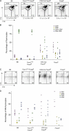
Distorted thymopoiesis is dependent on Foxp3 loss in the T cell lineage. Thymopoiesis was assessed by examining total thymus cellularity and by flow cytometric analysis of thymocyte subsets. (A) Representative CD4 and CD8 thymocyte profiles for wild-type, Foxp3-deficient, Foxp3fl CD4-Cre, and Foxp3fl Foxn1Cre mice. Average thymus cellularity (mean ± SD) is shown below each graph. (B) Percentages of thymocytes in CD4CD8 DN, CD4+CD8+ DP, CD4+ single positive, and CD8+ single positive subsets. Each data point represents a single mouse. (C) Representative flow cytometric profiles of DN thymocyte subsets and (D) percentages of DN1, DN2, DN3, and DN4 subsets in wild-type, Foxp3-deficient, Foxp3fl CD4-Cre, and Foxp3fl Foxn1Cre mice.
Figure 4.
Figure 4.
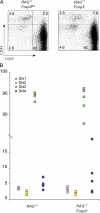
Foxp3 deficiency does not affect early thymopoiesis in disease-free Rag-deficient mice. (A) Representative flow cytometric analysis of DN1, DN2, DN3, and DN4 thymocyte subsets and (B) their percentages in Rag2 / and Foxp3-deficient Rag2 / mice. Each point represents a single mouse (n = 5). No statistically significant differences were observed between two groups of mice.

References

    1. Wildin, R.S., S. Smyk-Pearson, and A.H. Filipovich. 2002. Clinical and molecular features of the immunodysregulation, polyendocrinopathy, enteropathy, X linked (IPEX) syndrome. J. Med. Genet. 39:537–545. - PMC - PubMed
    1. Godfrey, V.L., J.E. Wilkinson, E.M. Rinchik, and L.B. Russell. 1991. Fatal lymphoreticular disease in the scurfy (sf) mouse requires T cells that mature in a sf thymic environment: potential model for thymic education. Proc. Natl. Acad. Sci. USA. 88:5528–5532. - PMC - PubMed
    1. Bennett, C.L., and H.D. Ochs. 2001. IPEX is a unique X-linked syndrome characterized by immune dysfunction, polyendocrinopathy, enteropathy, and a variety of autoimmune phenomena. Curr. Opin. Pediatr. 13:533–538. - PubMed
    1. Ferguson, P.J., S.H. Blanton, F.T. Saulsbury, M.J. McDuffie, V. Lemahieu, J.M. Gastier, U. Francke, S.M. Borowitz, J.L. Sutphen, and T.E. Kelly. 2000. Manifestations and linkage analysis in X-linked autoimmunity-immunodeficiency syndrome. Am. J. Med. Genet. 90:390–397. - PubMed
    1. Godfrey, V.L., J.E. Wilkinson, and L.B. Russell. 1991. X-linked lymphoreticular disease in the scurfy (sf) mutant mouse. Am. J. Pathol. 138:1379–1387. - PMC - PubMed

Publication types

MeSH terms

Substances