. 2007 Aug 16;26(38):5553-63.
doi: 10.1038/sj.onc.1210351. Epub 2007 Mar 12.

Cancer cells express aberrant DNMT3B transcripts encoding truncated proteins

Affiliations

Cancer cells express aberrant DNMT3B transcripts encoding truncated proteins

K R Ostler et al. Oncogene. .

Abstract

Cancer cells display an altered distribution of DNA methylation relative to normal cells. Certain tumor suppressor gene promoters are hypermethylated and transcriptionally inactivated, whereas repetitive DNA is hypomethylated and transcriptionally active. Little is understood about how the abnormal DNA methylation patterns of cancer cells are established and maintained. Here, we identify over 20 DNMT3B transcripts from many cancer cell lines and primary acute leukemia cells that contain aberrant splicing at the 5' end of the gene, encoding truncated proteins lacking the C-terminal catalytic domain. Many of these aberrant transcripts retain intron sequences. Although the aberrant transcripts represent a minority of the DNMT3B transcripts present, Western blot analysis demonstrates truncated DNMT3B isoforms in the nuclear protein extracts of cancer cells. To test if expression of a truncated DNMT3B protein could alter the DNA methylation patterns within cells, we expressed DNMT3B7, the most frequently expressed aberrant transcript, in 293 cells. DNMT3B7-expressing 293 cells have altered gene expression as identified by microarray analysis. Some of these changes in gene expression correlate with altered DNA methylation of corresponding CpG islands. These results suggest that truncated DNMT3B proteins could play a role in the abnormal distribution of DNA methylation found in cancer cells.


Figures

Figure 1
Figure 1
Cancer cells express aberrant DNMT3B transcripts as demonstrated by reverse-transcription PCR (RT–PCR). (a) DNMT3B cDNA was amplified from exon 9 to exon 13 in several cancer cell lines. DNA sizing is shown at the left. Product A is derived from a normal DNMT3B transcript that lacks exon 10, either DNMT3B2 or DNMT3B3. Product B is derived from DNMT3B1, which contains exon 10. Product C is an abnormally migrating species, the DNMT3B7 transcript. Amplification of the GAPD cDNA served as a loading control, demonstrating equal amounts of input cDNA from each cDNA source (bottom panel). (b) Alternative splicing of the DNMT3B gene. The protein-encoding exons of the DNMT3B gene are indicated at the top of the figure. Six major splice forms of the DNMT3B gene have been described, DNMT3B1-6. The exons contained in a particular transcript are shown in solid black rectangles. Exons excluded through alternative splicing are indicated by rectangular outlines. Premature translational stop codons are indicated by stop signs. The structure of the 5′ ends of the DNMT3B4 and DNMT3B5 transcripts are not known (Robertson et al., 1999). The most widely expressed aberrant DNMT3B transcript in cancer cells, DNMT3B7, is indicated in the middle of the figure. The retained intron sequence is indicated with a solid gray rectangle. The ΔDNMT3B1-4 transcripts identified recently in non-small-cell lung cancer differ from each other with respect to alternative splicing of exons 7 and 10 (numbering according to exons in DNMT3B1-3 transcripts), and ΔDNMT3B5-6 differ from each other with respect to alternative splicing of exon 7, indicated by hatched bars (Wang et al., 2006a, b).
Figure 2
Figure 2
Identification of truncated DNMT3B proteins by Western blotting. (a) Identification of truncated DNMT3B proteins in extracts from cancer cell lines. Upper panel, cytosolic and nuclear extracts (60 μg each) from SK-BR-3 and HeLa were probed for DNMT3B by Western blotting. The positions of full-length DNMT3B and truncated DNMT3B7 are indicated to the right. The positions of the molecular weight markers are given at the left. C, cytoplasmic extract; N, nuclear extract. Middle panel, parallel blot to that shown in the upper panel probed with the same antibody plus the antigenic peptide. Bottom panel, equal loading of cytosolic extracts and integrity of nuclear extracts as demonstrated by Western blotting using GAPDH. (b) Expression of DNMT3B7 in stable 293 cell lines. Upper panel, cytosolic and nuclear extracts (40–50 μg each) from vector-transfected or DNMT3B7-transfected293 cells demonstrate expression of DNMT3B7 in the nuclear fraction. Middle panel, parallel blot to that shown in the top panel probed with the same antibody plus the antigenic peptide. Bottom panel, equal loading of cytosolic extracts and integrity of nuclear extracts as demonstrated by Western blotting using GAPDH.
Figure 3
Figure 3
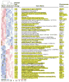
Gene expression changes in DNMT3B7-expressing 293 cells. A heat map shows the 51 genes whose expression changed with DNMT3B7 expression (red, overexpression; blue, under expression). Four samples of vector-transfected cells were compared to three samples each of DNMT3B7-expressing cells, and the average-fold change for each probe is listed. Gene names are in bold for the genes whose expression changes were validated by semiquantitative RT–PCR (data not shown). The chromosomal locus of each gene is given at the right, and the genes located on chromosomes 1, 9 and16, and the X chromosome are indicated by highlighting. Some genes (e.g., PRKCB1 and MID1) are listed more than once in the figure, because the microarray contained multiple probes for these genes.
Figure 4
Figure 4
293 cells overexpressing DNMT3B7 demonstrate gene expression changes that correspond with altered DNA methylation within some CpG islands as determined by sodium bisulfite analysis. Methylated CpG dinucleotides are represented by filled-in black circles, and unmethylated CpG dinucleotides are represented by open circles. Each numbered row represents an individual clone, and the CpG dinucleotide number is given across the top of each section. The number of identical clones is given in parentheses after a representative row. (a) Analysis of the methylation state of 18 individual CpG dinucleotides from the portion of the CDH1 CpG island that is located just 5′ to the gene’s transcriptional start. Hypermethylation of particular CpG dinucleotides in both line 1 and line 2 were statistically significant and are indicated by daggers. (b) Analysis of the methylation state of 12 individual CpG dinucleotides from the portion of the MAGEA3 CpG island that is located overlapping with the gene’s transcriptional start. (c) Analysis of the methylation state of two portions of the PLP2 CpG island. At the left, the figure shows the methylation state of 19 individual CpG dinucleotides from a part of the CpG island located just 5′ to the gene’s transcriptional start, and at the right, the figure indicates the methylation state of 24 additional CpG dinucleotides located just 3′ to the translational start. Hypomethylation of one particular CpG residue in the 3′ portion of the CpG island is indicated with an asterisk. (d) Analysis of the methylation state of seven individual CpG dinucleotides from the portion of the SH2D1A CpG island that is located within exon 2.

References

    1. Aggerholm A, Holm MS, Guldberg P, Olesen LH, Hokland P. Promoter hypermethylation of p15INK4B, HIC1, CDH1, and ER is frequent in myelodysplastic syndrome and predicts poor prognosis in early-stage patients. Eur J Haematol. 2006;76:23–32. - PubMed
    1. Bachman KE, Park BH, Rhee I, Rajagopalan H, Herman JG, Baylin SB, et al. Histone modifications and silencing prior to DNA methylation of a tumor suppressor gene. Cancer Cell. 2003;3:89–95. - PubMed
    1. Bartel F, Taubert H, Harris LC. Alternative and aberrant splicing of MDM2 mRNA in human cancer. Cancer Cell. 2002;2:9–15. - PubMed
    1. Beaulieu N, Morin S, Chute IC, Robert M-F, Nguyen H, MacLeod AR. An essential role for DNA methyltransferase DNMT3B in cancer cell survival. J Biol Chem. 2002;277:28176–28181. - PubMed
    1. Bestor TH. The DNA methyltransferases of mammals. Hum Molec Genet. 2000;9:2395–2402. - PubMed

Publication types

MeSH terms

Substances