

Prognostic significance of vascular endothelial cell growth factors -A, -C and -D in breast cancer and their relationship with angio- and lymphangiogenesis
- PMID: 17353919
- PMCID: PMC2360132
- DOI: 10.1038/sj.bjc.6603678
Prognostic significance of vascular endothelial cell growth factors -A, -C and -D in breast cancer and their relationship with angio- and lymphangiogenesis
Abstract
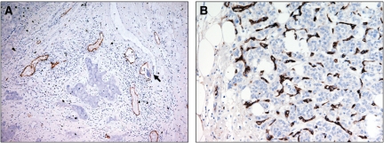
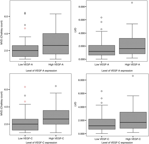
Vascular endothelial cell growth factors (VEGF)-A, -C and -D have potent angio and lymphangiogenic functions in experimental models, although their role in the progression of human breast cancer is unclear. The aims of the current study were to examine the relationship between the expression of the aforementioned growth factors with the angio and lymphangiogenic characteristics of breast cancer, and to assess their suitability as potential prognostic factors. Paraffin-embedded sections of 177 primary invasive breast cancer, with complete clinical follow-up information for 10 years, were stained for VEGF-A, -C, -D, podoplanin and CD34 using standard immunohistochemical approaches. The expression of the growth factors was correlated with clinicopathological criteria and patients' survival. Lymph vessel density (LVD) and microvessel density (MVD) were assessed and correlated with expression of the growth factors. Vascular endothelial cell growth factor-A, -C and -D were highly expressed in 40, 37 and 42% of specimens, respectively. High expression of VEGF-A and - C, but not of -D, was associated with a higher LVD (P=0.013 and P=0.014, respectively), a higher MVD (P<0.001 and P=0.002, respectively), the presence of lymph node metastasis (P<0.001 and P<0.001, respectively), distant metastasis (P=0.010 and P=0.008, respectively) and a shorter Overall Survival (P=0.029 and 0.028, respectively). In conclusion, breast cancers that express high levels of VEGF-A and -C are characterised by a poor prognosis, likely through the induction of angio and lymphangiogenesis. Examination of expression of VEGF-A and -C in breast cancer may be beneficial in the identification of a subset of tumours that have a higher probability of recurrence and metastatic spread.
Figures


References
-
- Bevilacqua P, Barbareschi M, Verderio P, Boracchi P, Caffo O, Dalla Palma P, Meli S, Weidner N, Gasparini G (1995) Prognostic value of intratumoral microvessel density, a measure of tumor angiogenesis, in node-negative breast carcinoma – results of a multiparametric study. Breast Cancer Res Treat 36: 205–217 - PubMed
-
- Choi WW, Lewis MM, Lawson D, Yin-Goen Q, Birdsong GG, Cotsonis GA, Cohen C, Young AN (2005) Angiogenic and lymphangiogenic microvessel density in breast carcinoma: correlation with clinicopathologic parameters and VEGF-family gene expression. Mod Pathol 18: 143–152 - PubMed
-
- Currie MJ, Hanrahan V, Gunningham SP, Morrin HR, Frampton C, Han C, Robinson BA, Fox SB (2004) Expression of vascular endothelial growth factor D is associated with hypoxia inducible factor (HIF-1alpha) and the HIF-1alpha target gene DEC1, but not lymph node metastasis in primary human breast carcinomas. J Clin Pathol 57: 829–834 - PMC - PubMed
MeSH terms
Substances
LinkOut - more resources
Full Text Sources
Other Literature Sources
Medical
