. 2007 Apr;5(4):e79.
doi: 10.1371/journal.pbio.0050079.

An aromatic sensor with aversion to damaged strands confers versatility to DNA repair

Affiliations

An aromatic sensor with aversion to damaged strands confers versatility to DNA repair

Olivier Maillard et al. PLoS Biol. 2007 Apr.

Abstract

It was not known how xeroderma pigmentosum group C (XPC) protein, the primary initiator of global nucleotide excision repair, achieves its outstanding substrate versatility. Here, we analyzed the molecular pathology of a unique Trp690Ser substitution, which is the only reported missense mutation in xeroderma patients mapping to the evolutionary conserved region of XPC protein. The function of this critical residue and neighboring conserved aromatics was tested by site-directed mutagenesis followed by screening for excision activity and DNA binding. This comparison demonstrated that Trp690 and Phe733 drive the preferential recruitment of XPC protein to repair substrates by mediating an exquisite affinity for single-stranded sites. Such a dual deployment of aromatic side chains is the distinctive feature of functional oligonucleotide/oligosaccharide-binding folds and, indeed, sequence homologies with replication protein A and breast cancer susceptibility 2 protein indicate that XPC displays a monomeric variant of this recurrent interaction motif. An aversion to associate with damaged oligonucleotides implies that XPC protein avoids direct contacts with base adducts. These results reveal for the first time, to our knowledge, an entirely inverted mechanism of substrate recognition that relies on the detection of single-stranded configurations in the undamaged complementary sequence of the double helix.


Conflict of interest statement

Competing interests. The authors have declared that no competing interests exist.

Figures

Figure 1
Figure 1. Evolutionary Conserved Residues in the Proposed DNA-Binding Domain of XPC Protein
Sequence comparison between eukaryotic XPC homologs. There are two homologous genes in S. pombe. Amino acids targeted by site-directed mutagenesis are highlighted. Y, F, W, aromatic (orange); K, L, positively charged (green); P, H, S, other highly conserved positions (yellow).
Figure 2
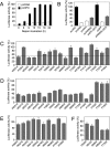
Figure 2. Screening for Repair-Deficient XPC Mutants
(A) Time course of host cell reactivation assay. Human XP–C fibroblasts were transfected with an expression vector coding for wild-type XPC (pcXPC) or the empty control vector (pcDNA). Repair complementation was assessed after the indicated times by monitoring luciferase expression from a UV-irradiated reporter construct. Excision is reported as the percentage of wild-type activity after 15 h of incubation (± SD). (B) Specificity of the repair assay. XP–C fibroblasts were transfected with expression vectors coding for wild-type XPC (pcXPC or pXPC–GFP), wild-type XPA (pcXPA), the control vectors (pcDNA and pGFP), or vectors containing the Trp690Ser mutant sequences (pcW690S or pW690S–GFP). Excision is reported as the percentage of wild-type activity (15-h incubations). (C) Deletion of aromatic side chains. XP–C fibroblasts were transfected with vector pcXPC carrying the indicated mutations. DNA repair is expressed as the percentage of wild-type complementation (15-h incubations) after deduction of background luciferase activity obtained with the control vector. The dashed line indicates a threshold of 50% reduction in repair activity. (D) Replacement of aromatic residues by amino acids of different properties. (E) Deletion of the positively charged side chains of conserved Lys or Arg residues. (F) Ala substitutions of absolutely conserved positions in the center of the DNA-binding domain.
Figure 3
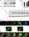
Figure 3. Normal Cellular Expression and Localization of Repair-Deficient XPC Mutants
(A) Immunoblot analysis of XP–C fibroblasts transfected with pcXPC vectors coding for wild-type protein or repair-deficient mutants. Soluble cell lysates (20 μg) were separated on polyacrylamide gels and probed for XPC protein using a specific monoclonal antibody directed against the C-terminal sequence of human XPC. Glyceraldehyde-3-phosphate dehydrogenase (GAPDH) was recorded as the internal standard. Lane 2: XP–C fibroblasts transfected with the pcDNA control vector. (B) Densitometric quantification of three to five independent experiments. The intensity of immunoreactive bands corresponding to XPC protein was normalized against the glyceraldehyde-3-phosphate dehydrogenase standard and reported as the percentage of the wild-type signal (± SD). (C) Immunoblot analysis of XP–C fibroblasts transfected with pGFP (lane 1), or the vectors coding for wild-type (lane 2) or mutant XPC proteins (lanes 3–7) fused to GFP. The primary antibody was directed against GFP. (D) Time course of fluorescent fusion protein expression in XP–C fibroblasts transfected with pXPC–GFP containing the wild-type sequence. Nuclei were stained with Hoechst reagent. (E) Distribution of GFP in XP–C fibroblasts. (F and G) Representative images demonstrating the nuclear localization of repair-deficient mutants 15 h after transfection.
Figure 4
Figure 4. Affinity of XPC Protein for Native Single-Stranded Oligonucleotides
(A) Analysis of MBP–XPC fusion protein by Coomassie staining of a denaturing 8% polyacrylamide gel. Lane 1, markers; lane 2, purified fraction. (B) Electrophoretic mobility shift assay demonstrating the preference of wild-type XPC protein for UV-irradiated duplexes over the unirradiated control (lane 1). Radiolabeled double-stranded DNA fragments of 65 base pairs (2 nM) were incubated for 30 min with the MBP–XPC fusion product (50 nM) and duplex poly[dI-dC] (10 ng/μl). F, free DNA; B, protein-bound DNA. (C) Preference of XPC protein for binding to single-stranded 65-mer oligonucleotides (lanes 1–4) relative to undamaged 65-mer duplexes (lanes 5–8). (D) Competition with single-stranded DNA. Radiolabeled 65-mer duplexes were UV-irradiated (1.8 kJ/m2) and incubated at a concentration of 2 nM with XPC protein (50 nM), increasing amounts of unlabeled single-stranded oligomers of 65 nucleotides, and duplex poly[dI-dC] (10 ng/μl). The fractions of protein-bound oligomers were determined by electrophoretic mobility shift assay, quantified by laser scanning densitometry, and expressed as the percentage of binding observed in the absence of competitor DNA (± SD). (E) Competition with double-stranded DNA. Radiolabeled 65-mer oligonucleotides (2 nM) were incubated with XPC protein (50 nM), 100 ng of plasmid DNA (pcDNA) exposed to the indicated UV doses, and duplex poly[dI-dC] (10 ng/μl). The fractions of protein-bound oligomers were quantified and expressed as the percentage of binding determined in the presence of undamaged competitor DNA (± SD). (F) Suppression of single-stranded DNA binding by UV irradiation. Radiolabeled 65-mer oligonucleotides (2 nM), exposed to the indicated UV doses, were incubated for 30 min with XPC protein (100 nM) and duplex poly[dI-dC] (10 ng/μl). Lane 1: no XPC protein. (G) Quantification by laser scanning densitometry of two independent experiments performed with UV-irradiated single-stranded oligonucleotides (± SD).
Figure 5
Figure 5. The Trp690Ser Mutant Is Defective in Substrate Binding
(A) Pull-down assays were performed by coincubating Sf9 cell lysate (5 μl) containing wild-type XPC protein and 50 μl of single-stranded DNA beads. The binding buffer was supplemented with the indicated concentrations of NaCl. The fractions of free (F) and bound (B) protein were separated and analyzed by gel electrophoresis and immunobloting using specific monoclonal antibodies. The panel on the right provides a quantitative evaluation of three independent binding assays showing the proportion of pulled-down XPC protein at the different ionic strengths. (B) Pull-down assay with Sf9 cell lysate (5 μl) containing the Trp690Ser mutant (left) and quantitative evaluation of three independent experiments (right).
Figure 6
Figure 6. DNA-Binding Deficiency of Trp690 and Phe733 Mutants
(A) Pull-down assays were performed by coincubating Sf9 cell lysate (5 μl) containing wild-type XPC or the indicated Ala mutants and single-stranded DNA beads. The binding buffer contained 0.3 M NaCl. The fractions of free (F) and bound (B) protein were separated and analyzed by gel electrophoresis and immunobloting using specific monoclonal antibodies. The panel on the right shows the quantitative evaluation of three independent binding assays (± SD). (B) Side-by-side comparison of the DNA-binding capacity of wild-type XPC protein (lanes 7 and 8) and the indicated mutants (lanes 1–6).
Figure 7
Figure 7. Single-Stranded Oligonucleotide-Binding Defect
(A) Analysis of immunoprecipitated XPC by Coomassie staining of a denaturing 8% polyacrylamide gel. The MBP–XPC fusions were purified from Sf9 lysates using monoclonal anti-MBP antibodies linked to paramagnetic beads. Lane 1, markers; lane 2, wild-type MBP–XPC; lane 3, fusion protein containing the Trp690Ser reference mutation. (B) Binding of wild-type XPC and Trp690Ser mutant to single-stranded oligonucleotides. Immunoprecipitated MBP–XPC protein (100 ng, 3 nM) was incubated with 32P-labeled 65-mer oligonucleotides (2 nM). The DNA molecules captured by XPC protein were separated from the free oligonucleotides and quantified in a scintillation counter. Single-stranded DNA-binding activity (± SD) is reported as the radioactivity immobilized by XPC after deduction of the background binding determined with empty beads. (C) Differential binding to distinct DNA conformations. Immunoprecipitated MBP–XPC protein (100 ng, 3 nM) was incubated with 32P-labeled substrates (2 nM) consisting of 65-mer homoduplexes, 65-mer heteroduplexes with a central 3-nucleotide bubble, or 65-mer single-stranded oligonucleotides. The DNA molecules captured by XPC protein were separated from free DNA and quantified in a scintillation counter. DNA-binding activity (mean values of two experiments) is reported as the radioactivity immobilized by XPC after deduction of the background binding determined with empty beads. (D) Comparison between wild-type XPC and Ala mutants. Paramagnetic beads containing the indicated amounts of immunopurified MBP–XPC protein were incubated with 32P-labeled 65-mer oligonucleotides (2 nM). DNA associated with XPC protein was separated from the free oligonucleotides and quantified in a scintillation counter. Single-stranded DNA binding activity (mean values of four experiments) is reported as the radioactivity immobilized by XPC after deduction of the background binding to empty beads.
Figure 8
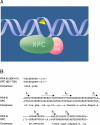
Figure 8. Versatile Damage Recognition: Detection of Single-Stranded Configurations in the Undamaged Strand of the Double Helix
(A) Initial interaction of XPC protein with damaged sites driven by an affinity for native single-stranded DNA. The triangle symbolizes a helix-distorting bulky lesion. This mechanism with inverted DNA strand specificity directs XPC protein to the undamaged strand and the downstream factors of the GGR pathway to the damaged strand. (B) Alignment of the RPA-B and XPC DNA-binding sequences. The consensus was derived using the following amino acid classes [47]: hydrophobic (h, ALICVMYFW); the aliphatic subset of these (a, ALIVMC); small (s, ACDGNPSTV); the “tiny” subset of these (u, GAS); polar (p, CDEHKNQRST); charged (c, DEHKR), positively charged (+, HKR); and negatively charged (n, DE). The length of nonalignable gaps is indicated in parentheses and the β-sheet elements are indicated by the arrows.

Similar articles

Cited by

References

    1. Sancar A. DNA excision repair. Annu Rev Biochem. 1996;65:43–81. - PubMed
    1. Wood RD. Nucleotide excision repair in mammalian cells. J Biol Chem. 1997;272:23465–23468. - PubMed
    1. De Laat WL, Jaspers NG, Hoeijmakers JHJ. Molecular mechanism of nucleotide excision repair. Genes Dev. 1999;13:768–785. - PubMed
    1. Gillet LC, Schärer OD. Molecular mechanisms of mammalian global genome nucleotide excision repair. Chem Rev. 2006;106:253–276. - PubMed
    1. Satoh MS, Jones CJ, Wood RD, Lindahl T. DNA excision-repair defect of xeroderma pigmentosum prevents removal of a class of oxygen free radical-induced base lesions. Proc Natl Acad Sci U S A. 1993;90:6335–6339. - PMC - PubMed

Publication types

LinkOut - more resources