. 2007 May;98(5):674-84.
doi: 10.1111/j.1349-7006.2007.00440.x. Epub 2007 Mar 9.

Inhibition of ADAM17 reduces hypoxia-induced brain tumor cell invasiveness

Affiliations

Inhibition of ADAM17 reduces hypoxia-induced brain tumor cell invasiveness

Xuguang Zheng et al. Cancer Sci. 2007 May.

Abstract

The membrane-anchored metalloproteinase tumor necrosis factor-alpha-converting enzyme (TACE/a disintegrin and metalloproteinase [ADAM] 17) is key in proteolytic ectodomain shedding of several membrane-bound growth factors, cytokines and receptors. The expression and activity of ADAM17 increases under some pathological conditions including stroke, and promotes neural progenitor cell migration and contributes to stroke-induced neurogenesis. Hypoxia initiates cellular invasive processes that occur under both physiological and pathological conditions such as invasion and metastasis of some tumors. In the present study, we sought to elucidate whether ADAM17 contributes to brain tumor invasion. To this end, we examined the role of ADAM17 in the invasiveness of two different brain tumor cell lines, 9L rat gliosarcoma and U87 human glioma, under normoxic and hypoxic conditions. Additionally, we tested the effects of ADAM17 suppression on in vitro tumor cell invasion by means of ADAM17 proteolytic inhibitors and specific small interfering RNA. We found that tumor cells upregulated ADAM17 expression under hypoxia, and that ADAM17 activity correlated with increased tumor cell invasion. Conversely, suppression of ADAM17 proteolysis decreased invasiveness induced by hypoxia in 9L and U87 cells. Furthermore, the contribution of ADAM17 to tumor invasion was independent of matrix metalloproteinase (MMP)-2 and MMP-9 activity. ADAM17 was also found to activate the epidermal growth factor/phosphoinositide-3 kinase/serine/threonine kinase signal transduction pathway. Our data suggest that hypoxia-induced ADAM17 contributes to glioma cell invasiveness through activation of the EGFR signal pathway.


Figures
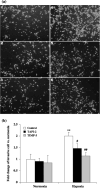
Figure 1
Figure 1
Effect of the a disintegrin and metalloproteinase (ADAM) 17 inhibitors tumor necrosis factor‐α protease inhibitor (TAPI)‐2 and tissue inhibitor of metalloproteinase (TIMP)‐3 on the in vitro invasiveness of 9L brain tumor cells cultured under normoxic (20% O2) or hypoxic (1% O2) conditions. (a) Representative pictures of 9L cell invasiveness assay with different treatments. 1, Normoxic control; 2, normoxia + 20 µmol/L TAPI‐2; 3, normoxia + 40 ng/mL TIMP‐3; 4, hypoxia; 5, hypoxia + 20 µmol/L TAPI‐2; 6, hypoxia + 40 ng/mL TIMP‐3. (b) Relative invasive cell number compared to normoxia control. Data are shown as percentage of results under normoxia. The values indicated by two asterisks (**) were significantly different (P < 0.01) from normoxia; the values indicated by hatches (# P < 0.05; ## P < 0.01) were significantly different from the hypoxic control.
Figure 2
Figure 2
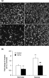
Effect of the a disintegrin and metalloproteinase (ADAM) 17 inhibitor tumor necrosis factor‐α protease inhibitor (TAPI)‐2 on the in vitro invasiveness of U87 brain tumor cells cultured under normoxic (20% O2) or hypoxic (1% O2) conditions. (a) Representative pictures of U87 cell invasiveness assay with different treatments. 1, Normoxic control; 2, normoxia + 20 µmol/L TAPI‐2; 3, hypoxia; 4, hypoxia + 20 µmol/L TAPI‐2. (b) Relative invasive cell number compared to normoxic control. Data are shown as percentage of results under normoxia. The values indicated by two asterisks (**) were significantly different (P < 0.01) from normoxia; the values indicated by hatches (# P < 0.01) were significantly different from hypoxic control.
Figure 3
Figure 3
Real‐time reverse transcription–polymerase chain reaction for hypoxia inducible factor (HIF)‐1α, a disintegrin and metalloproteinase (ADAM) 17, matrix metalloproteinase (MMP)‐2 and MMP‐9 at the RNA level in (a) 9L, (b) HIF‐1α, (c) ADAM17, (d) MMP‐2 and (e) MMP‐9. 9L cells under normoxic and hypoxic conditions for 12 h. RNA levels of HIF‐1α, ADAM17, MMP‐2 and MMP‐9 in 9L were all significantly increased under hypoxic conditions. In normoxic conditions, RNA levels of HIF‐1α, ADAM17, MMP‐2 and MMP‐9 were not significantly altered by incubation with ADAM17 inhibitors tumor necrosis factor‐α protease inhibitor (TAPI)‐2 or tissue inhibitor of metalloproteinase (TIMP)‐3. Under hypoxic conditions for 12 h, RNA levels of HIF‐1α, ADAM17, MMP‐2 and MMP‐9 in 9L were not significantly altered by incubation with ADAM17 inhibitors TAPI‐2 or TIMP‐3, compared to the hypoxic control. The values indicated by two asterisks (**) were significantly different (P < 0.01) from the normoxic control.
Figure 4
Figure 4
Western blot analysis for a disintegrin and metalloproteinase (ADAM) 17, matrix metalloproteinase (MMP)‐2 and MMP‐9 protein levels in 9L cells. 9L cells under normoxic and hypoxic conditions for 24 h. Protein levels of ADAM17, MMP‐2 and MMP‐9 in 9L were all significantly increased under hypoxia. In normoxic conditions, protein levels of ADAM17, MMP‐2 and MMP‐9 in 9L were not significantly altered by incubation with the ADAM17 inhibitor tumor necrosis factor‐α protease inhibitor (TAPI)‐2; protein levels of ADAM17 and MMP‐2 in 9L were not significantly altered by incubation with the ADAM17 inhibitor tissue inhibitor of metalloproteinase (TIMP)‐3. Meanwhile, expression of MMP‐9 in 9L cells treated with TIMP‐3 for 24 h increased compared to the normoxic control. In hypoxic conditions, protein levels of ADAM17, MMP‐2 and MMP‐9 in 9L were not significantly altered by incubation with ADAM17 inhibitors TAPI‐2 or TIMP‐3 for 24 h, compared to hypoxic control.
Figure 5
Figure 5
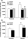
Effect of a disintegrin and metalloproteinase (ADAM) 17 inhibition on hypoxia‐induced ADAM17 activity of (a) 9L and (b) U87 cells. (a) Hypoxic treatment of 9L for 24 h increased the level of ADAM17 activity by 38.7% (P < 0.01). Treatment with ADAM17 inhibitors tumor necrosis factor‐α protease inhibitor (TAPI)‐2 or tissue inhibitor of metalloproteinase (TIMP)‐3 decreased ADAM17 by 40.7% (P < 0.01) and 30.9% (P < 0.01), respectively, in hypoxic conditions, compared to the hypoxic control. 20 µM TAPI‐2 decreased ADAM17 activity by 16.8% (P < 0.05) in normoxic conditions. (b) Hypoxic treatment of U87 for 24 h increased the level of ADAM17 activity by 46.9% (P < 0.01). Treatment with ADAM17 inhibitor TAPI‐2 (20 µmol/L) decreased ADAM17 activity by 21.3% (P < 0.05) in normoxic conditions, compared to the normoxic control; treatment with TAPI‐2 (20 µmol/L) decreased ADAM17 activity by 25.8% (P < 0.01) in hypoxic conditions, compared to the hypoxic control. The values indicated by asterisks were significantly different (*P < 0.05; **P < 0.01) from normoxic control; the values indicated by hatches (### P < 0.01) were significantly different from hypoxic control.
Figure 6
Figure 6
Effect of a disintegrin and metalloproteinase (ADAM) 17 inhibition on hypoxia‐induced matrix metalloproteinase (MMP) activity of 9L cells. (a) Zymogram analysis of MMP‐9 and MMP‐2 secreted by 9L cells. Cell lysates (upper) and supernatants (lower) were analyzed in normoxic conditions and hypoxic conditions. Pictures are representative of three independent experiments. (b–d) The density of each band was measured using Gelpro 4.5 software. Hypoxia resulted in a 1.50‐fold increase in cell lysate MMP‐2 activity (P < 0.05); 20 µmol/L tumor necrosis factor‐α protease inhibitor (TAPI)‐2 and 40 ng/mL tissue inhibitor of metalloproteinase (TIMP)‐3 did not alter MMP‐2 levels (b). At the same time, hypoxia increased 9L cell supernatant (c) MMP‐9 and (d) MMP‐2 activities 1.40‐fold and 1.50‐fold, respectively. 20 µmol/L TAPI‐2 did not alter (c) MMP‐9 or (d) MMP‐2 activities in either normoxic or hypoxic conditions. 40 ng/mL TIMP‐3 decreased (c) MMP‐9 and (d) MMP‐2 levels significantly in hypoxic conditions, when compared to the hypoxic control (e). The values indicated by two asterisks were significantly different (*P < 0.01) from normoxic control; the values indicated by hatches (# P < 0.05) were significantly different from hypoxic control.
Figure 7
Figure 7
Enzyme‐linked immunosorbent assay analysis for phospho‐extracellular signal‐regulated kinase (p‐ERK) and phospho‐serine/threonine kinase (p‐AKT) protein levels in 9L and U87 cells. 9L and U87 cells were cultured under normoxic and hypoxic conditions for 24 h. p‐AKT levels in both cell lines were significantly increased under hypoxic conditions, compared to normoxic conditions. Treatment with tumor necrosis factor‐α protease inhibitor (TAPI)‐2 (20 µmol/L) decreased p‐AKT level significantly in both (b) 9L (28.2% inhibition compared to hypoxia control) and (d) U87 (37.4% inhibition compared to hypoxia control) cell lines. However, hypoxia and treatment of TAPI‐2 did not alter the levels of p‐ERK2 in either (a) 9L or (c) U87 cell lines.
Figure 8
Figure 8
Effect of human a disintegrin and metalloproteinase (ADAM) 17 small interfering RNA (siRNA) transfection on ADAM17 mRNA expression and activity in U87 cells under normoxic and hypoxic conditions. ADAM17 mRNA and activity levels were significantly increased under hypoxic conditions compared to normoxic conditions. ADAM17 siRNA transfection reduced ADAM17 mRNA expression and activity in both normoxic and hypoxic conditions.
Figure 9
Figure 9
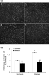
Effect of human a disintegrin and metalloproteinase (ADAM) 17 small interfering RNA (siRNA) transfection on the in vitro invasiveness of U87 brain tumor cells cultured under normoxic (20% O2) or hypoxic (1% O2) conditions. (a) Representative pictures of U87 cell invasion assay with different treatments. 1, Cells under normoxic conditions transfected with control siRNA; 2, cells under normoxic conditions transfected with ADAM17 siRNA; 3, cells under hypoxic conditions transfected with control siRNA; 4, cells under hypoxic conditions transfected with ADAM17 siRNA. (b) Relative invasive cell number compared to normoxic control. Data are shown as percentage of results under normoxic conditions. The values indicated by two asterisks (**) were significantly different (P < 0.01) from normoxia; the values indicated by hatches (# P < 0.01) were significantly different from the hypoxic control.
Figure 10
Figure 10
Western blot analysis for a disintegrin and metalloproteinase (ADAM) 17, epidermal growth factor receptor (EGFR), phosphorylated EGFR (p‐EGFR), serine/threonine kinase (AKT) and phosphorylated AKT (p‐AKT) protein levels in U87 cells after small interfering RNA (siRNA) transfection. U87 cells transfected with siRNA cultured under normoxic and hypoxic conditions for 24 h. Protein levels of ADAM17, p‐EGFR, and p‐AKT were all significantly increased under hypoxic conditions; protein levels of EGFR and AKT were not significantly altered by hypoxia. ADAM17 siRNA transfection decreased ADAM17, p‐EGFR and p‐AKT protein levels in both normoxic and hypoxic conditions. EGFR and AKT protein levels were not significantly altered by ADAM17 siRNA in either normoxic or hypoxic conditions.
Figure 11
Figure 11
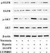
Effects of AG1478 and LY294002 on the epidermal growth factor receptor (EGFR), phosphorylated EGFR (p‐EGFR), serine/threonine kinase (AKT) and phosphorylated AKT (p‐AKT) protein levels in U87 cells under normoxic and hypoxic conditions. U87 cells were treated with EGFR activation inhibitor AG1478 and AKT activation inhibitor LY294002 under normoxic and hypoxic conditions. Protein levels of p‐EGFR and p‐AKT were all significantly increased under hypoxic conditions; protein levels of EGFR and AKT were not significantly altered by hypoxic treatment. AG1478 decreased p‐EGFR and p‐AKT protein levels in both normoxic and hypoxic conditions; EGFR and AKT protein levels were not significantly altered by AG1478 in either normoxic or hypoxic conditions. LY294002 decreased p‐AKT protein levels in both normoxic and hypoxic conditions; p‐EGFR, EGFR and AKT protein levels were not significantly altered by LY294002 in either normoxic or hypoxic conditions.
Figure 12
Figure 12
Effect of AG1478 and LY294002 on the in vitro invasiveness of U87 brain tumor cells cultured under normoxic or hypoxic conditions. (a) Representative pictures of U87 cell invasion assay with different treatments. 1, Normoxic control; 2, normoxia + 10 µmol/L AG1478; 3, normoxia + 20 µmol/L LY294002; 4, hypoxia; 5, hypoxia + 10 µmol/L AG1478; 6, hypoxia + 20 µmol/L LY294002. (b) Relative invasive cell number compared to normoxic control. Data are shown as percentage of results under normoxic conditions. The values indicated by two asterisks (**) were significantly different (P < 0.01) from normoxia; the values indicated by hatches (# P < 0.01) were significantly different from the hypoxic control.

Similar articles

Cited by

References

    1. Black RA. Tumor necrosis factor‐α converting enzyme. Int J Biochem Cell Biol 2002; 34: 1–5. - PubMed
    1. Black RA, Rauch CT, Kozlosky CJ et al. A metalloproteinase disintegrin that releases tumour‐necrosis factor‐α from cells. Nature 1997; 385: 729–33. - PubMed
    1. Buxbaum JD, Liu KN, Luo Y et al. Evidence that tumor necrosis factor alpha converting enzyme is involved in regulated alpha‐secretase cleavage of the Alzheimer amyloid protein precursor. J Biol Chem 1998; 273: 27 765–7. - PubMed
    1. Zatovicova M, Sedlakova O, Svastova E et al. Ectodomain shedding of the hypoxia‐induced carbonic anhydrase IX is a metalloprotease‐dependent process regulated by TACE/ADAM17. Br J Cancer 2005; 93: 1267–76. - PMC - PubMed
    1. Crowe PD, Walter BN, Mohler KM, Otten‐Evans C, Black RA, Ware CF. A metalloprotease inhibitor blocks shedding of the 80‐kD TNF receptor and TNF processing in T lymphocytes. J Exp Med 1995; 181: 1205–10. - PMC - PubMed

Publication types

MeSH terms